Natalia Kleinhans, Sarah F Larsen, Annette Estes, Elizabeth Aylward
{"title":"Intrinsic Functional Connectivity Alterations of the Fusiform Face Area in Autism Spectrum Disorder.","authors":"Natalia Kleinhans, Sarah F Larsen, Annette Estes, Elizabeth Aylward","doi":"10.3390/neurosci6020029","DOIUrl":null,"url":null,"abstract":"<p><p>Intrinsic connectivity of the fusiform face area (FFA) was assessed using resting-state functional magnetic resonance imaging (fMRI) to compare adults with autism spectrum disorder (ASD; <i>n</i> = 17) and age-, sex-, and IQ-matched typically developing controls (TD; <i>n</i> = 22). The FFA seed region was delineated in each participant using a functional localizer task. Whole brain analyses of FFA connectivity revealed increased connectivity between the right FFA and the vermis, sensorimotor cortex, and extended face-processing network in individuals with ASD compared to TD participants; the TD group did not demonstrate increased functional connectivity. No group differences were observed from the left FFA. The relationship between FFA connectivity and the ability to remember faces significantly differed between the groups. Better face memory performance was positively correlated with increased connectivity within general visual processing areas in the ASD participants; whereas for the TD group, better face memory performance was associated with increased connectivity with brain regions related to face encoding, recognition, and retrieval. FFA overconnectivity with face, emotion, and memory processing areas, along with atypical relationships between FFA-occipito-temporal connections and face memory performance highlights a possible mechanism underlying social dysfunction in individuals with ASD.</p>","PeriodicalId":74294,"journal":{"name":"NeuroSci","volume":"6 2","pages":""},"PeriodicalIF":2.0000,"publicationDate":"2025-04-01","publicationTypes":"Journal Article","fieldsOfStudy":null,"isOpenAccess":false,"openAccessPdf":"https://www.ncbi.nlm.nih.gov/pmc/articles/PMC12015912/pdf/","citationCount":"0","resultStr":null,"platform":"Semanticscholar","paperid":null,"PeriodicalName":"NeuroSci","FirstCategoryId":"1085","ListUrlMain":"https://doi.org/10.3390/neurosci6020029","RegionNum":0,"RegionCategory":null,"ArticlePicture":[],"TitleCN":null,"AbstractTextCN":null,"PMCID":null,"EPubDate":"","PubModel":"","JCR":"Q3","JCRName":"CLINICAL NEUROLOGY","Score":null,"Total":0}
引用次数: 0
Abstract
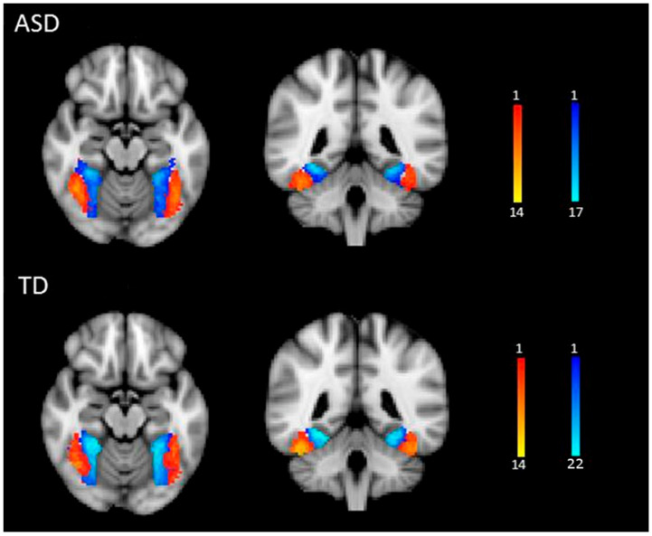
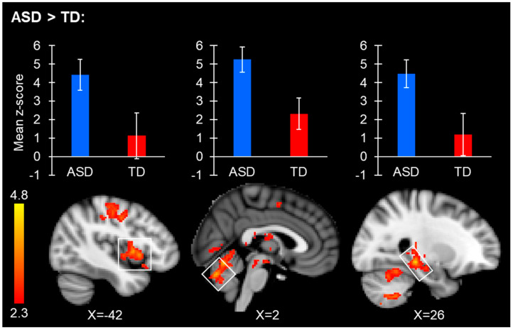
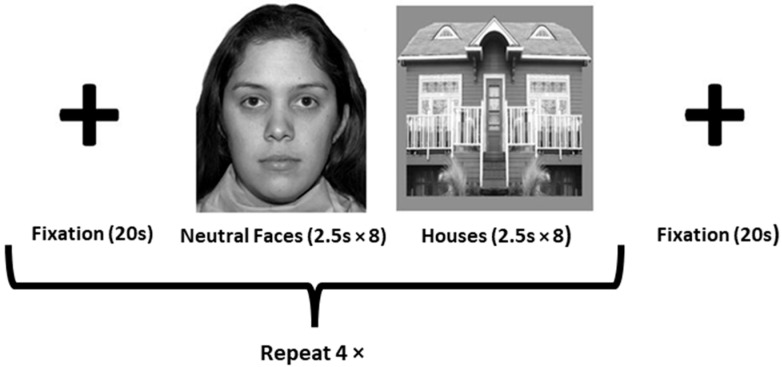
Intrinsic connectivity of the fusiform face area (FFA) was assessed using resting-state functional magnetic resonance imaging (fMRI) to compare adults with autism spectrum disorder (ASD; n = 17) and age-, sex-, and IQ-matched typically developing controls (TD; n = 22). The FFA seed region was delineated in each participant using a functional localizer task. Whole brain analyses of FFA connectivity revealed increased connectivity between the right FFA and the vermis, sensorimotor cortex, and extended face-processing network in individuals with ASD compared to TD participants; the TD group did not demonstrate increased functional connectivity. No group differences were observed from the left FFA. The relationship between FFA connectivity and the ability to remember faces significantly differed between the groups. Better face memory performance was positively correlated with increased connectivity within general visual processing areas in the ASD participants; whereas for the TD group, better face memory performance was associated with increased connectivity with brain regions related to face encoding, recognition, and retrieval. FFA overconnectivity with face, emotion, and memory processing areas, along with atypical relationships between FFA-occipito-temporal connections and face memory performance highlights a possible mechanism underlying social dysfunction in individuals with ASD.

 求助内容:
求助内容: 应助结果提醒方式:


