At the crossroad between Ewing sarcoma and neuroblastoma: a report of two cases of Ewing sarcoma with post treatment neuroblastoma-like differentiation.
{"title":"At the crossroad between Ewing sarcoma and neuroblastoma: a report of two cases of Ewing sarcoma with post treatment neuroblastoma-like differentiation.","authors":"Giorgia Arcovito, Giacomo Aringhieri, Virna Zampa, Luca Coccoli, Lorenzo Andreani, Alessandro Franchi","doi":"10.1186/s13000-025-01649-8","DOIUrl":null,"url":null,"abstract":"<p><strong>Background: </strong>Ewing sarcoma (ES) is the second most frequent sarcoma of bone, often affecting young patients and pursuing an aggressive clinical course. Among therapeutic choices, radio- and chemotherapy are employed in neoadjuvant setting, and they yield variable histological changes in neoplastic tissue, which mainly include necrosis and fibrosis. Cytodifferentiation is seldom observed in pediatric tumors such as nephroblastoma and rhabdomyosarcoma following treatment. Nevertheless, it represents an extremely rare phenomenon in ES.</p><p><strong>Case presentation: </strong>In this study we present the clinico-pathologic and molecular features of two cases of ES undergoing neuroblastoma-like differentiation after treatment. Both tumors were primarily located in bone and presented the histologic and immunohistochemical features of classic ES in needle biopsies. They were treated with standard chemotherapy protocols followed by surgical resection. The resection specimens of the primary tumor of patient 1 and pleural metastases of patient 2 presented foci of eosinophilic fibrillary stroma resembling neuropil and containing cellular elements with wide granular eosinophilic cytoplasm, eccentric nuclei containing vesicular chromatin and prominent nucleoli, reminiscent of ganglion cells. These cells were positive for chromogranin, synaptophysin and CD56, while CD99 was negative. Molecular confirmation of EWSR1 rearrangement was provided in both cases by next generation sequencing and FISH analysis.</p><p><strong>Conclusions: </strong>Evidence of neural differentiation in ES unravels interesting hints about its controversial histogenesis. Furthermore, awareness of this event must be increased to avoid misdiagnosis with neuroblastoma, which shows significant morphological overlap.</p>","PeriodicalId":11237,"journal":{"name":"Diagnostic Pathology","volume":"20 1","pages":"50"},"PeriodicalIF":2.3000,"publicationDate":"2025-04-23","publicationTypes":"Journal Article","fieldsOfStudy":null,"isOpenAccess":false,"openAccessPdf":"https://www.ncbi.nlm.nih.gov/pmc/articles/PMC12016484/pdf/","citationCount":"0","resultStr":null,"platform":"Semanticscholar","paperid":null,"PeriodicalName":"Diagnostic Pathology","FirstCategoryId":"3","ListUrlMain":"https://doi.org/10.1186/s13000-025-01649-8","RegionNum":3,"RegionCategory":"医学","ArticlePicture":[],"TitleCN":null,"AbstractTextCN":null,"PMCID":null,"EPubDate":"","PubModel":"","JCR":"Q2","JCRName":"PATHOLOGY","Score":null,"Total":0}
引用次数: 0
Abstract
Background: Ewing sarcoma (ES) is the second most frequent sarcoma of bone, often affecting young patients and pursuing an aggressive clinical course. Among therapeutic choices, radio- and chemotherapy are employed in neoadjuvant setting, and they yield variable histological changes in neoplastic tissue, which mainly include necrosis and fibrosis. Cytodifferentiation is seldom observed in pediatric tumors such as nephroblastoma and rhabdomyosarcoma following treatment. Nevertheless, it represents an extremely rare phenomenon in ES.
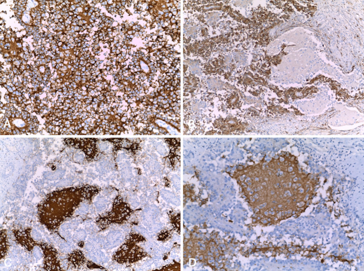
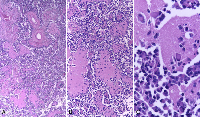
Case presentation: In this study we present the clinico-pathologic and molecular features of two cases of ES undergoing neuroblastoma-like differentiation after treatment. Both tumors were primarily located in bone and presented the histologic and immunohistochemical features of classic ES in needle biopsies. They were treated with standard chemotherapy protocols followed by surgical resection. The resection specimens of the primary tumor of patient 1 and pleural metastases of patient 2 presented foci of eosinophilic fibrillary stroma resembling neuropil and containing cellular elements with wide granular eosinophilic cytoplasm, eccentric nuclei containing vesicular chromatin and prominent nucleoli, reminiscent of ganglion cells. These cells were positive for chromogranin, synaptophysin and CD56, while CD99 was negative. Molecular confirmation of EWSR1 rearrangement was provided in both cases by next generation sequencing and FISH analysis.
Conclusions: Evidence of neural differentiation in ES unravels interesting hints about its controversial histogenesis. Furthermore, awareness of this event must be increased to avoid misdiagnosis with neuroblastoma, which shows significant morphological overlap.
期刊介绍:
Diagnostic Pathology is an open access, peer-reviewed, online journal that considers research in surgical and clinical pathology, immunology, and biology, with a special focus on cutting-edge approaches in diagnostic pathology and tissue-based therapy. The journal covers all aspects of surgical pathology, including classic diagnostic pathology, prognosis-related diagnosis (tumor stages, prognosis markers, such as MIB-percentage, hormone receptors, etc.), and therapy-related findings. The journal also focuses on the technological aspects of pathology, including molecular biology techniques, morphometry aspects (stereology, DNA analysis, syntactic structure analysis), communication aspects (telecommunication, virtual microscopy, virtual pathology institutions, etc.), and electronic education and quality assurance (for example interactive publication, on-line references with automated updating, etc.).

 求助内容:
求助内容: 应助结果提醒方式:


