{"title":"Pilomatricoma of the breast similar to breast cancer on ultrasound and elastography: a case report.","authors":"Shuanglian Zhu, Nianyu Xue","doi":"10.21037/acr-24-103","DOIUrl":null,"url":null,"abstract":"<p><strong>Background: </strong>Pilomatricoma, also known as calcifying epithelioma of Malherbe, is a benign tumor originating from human hair follicle stem cells and commonly observed in adolescents. Pilomatricomas can arise in any region with hair follicles, typically presenting as solitary lesions, predominantly affecting the facial region. Clinically, they manifest as firm, painless nodules, with both pathological and ultrasonographic findings often revealing calcifications. We report a rare case of pilomatricoma located on the breast, which exhibits characteristics are similar to breast cancer. Combining with ultrasound and elastography may provide some references for clinical diagnosis.</p><p><strong>Case description: </strong>We report a case of pilomatricoma located on the 47-year-old female breast, characterized by multiple punctate hyperechoic foci internally, presenting as a firm, non-tender mass on palpation and elastography. Without seeking treatment, she experienced pain in the lump two weeks before presentation. After applying an unspecified anti-inflammatory ointment, the lump markedly increased. Therefore, the rapid enlargement of the mass within a short period raised clinical suspicion of breast cancer, leading to surgical excision, which subsequently confirmed pilomatricoma through histopathological examination.</p><p><strong>Conclusions: </strong>Pilomatricomas occurring on the breast may present clinically and ultrasonographically similar to breast cancer, necessitating a focus on distinguishing between the two entities based on layer-derived differentiation on ultrasound and elastography.</p>","PeriodicalId":29752,"journal":{"name":"AME Case Reports","volume":"9 ","pages":"46"},"PeriodicalIF":0.7000,"publicationDate":"2025-03-11","publicationTypes":"Journal Article","fieldsOfStudy":null,"isOpenAccess":false,"openAccessPdf":"https://www.ncbi.nlm.nih.gov/pmc/articles/PMC12053875/pdf/","citationCount":"0","resultStr":null,"platform":"Semanticscholar","paperid":null,"PeriodicalName":"AME Case Reports","FirstCategoryId":"1085","ListUrlMain":"https://doi.org/10.21037/acr-24-103","RegionNum":0,"RegionCategory":null,"ArticlePicture":[],"TitleCN":null,"AbstractTextCN":null,"PMCID":null,"EPubDate":"2025/1/1 0:00:00","PubModel":"eCollection","JCR":"Q3","JCRName":"MEDICINE, GENERAL & INTERNAL","Score":null,"Total":0}
引用次数: 0
Abstract
Background: Pilomatricoma, also known as calcifying epithelioma of Malherbe, is a benign tumor originating from human hair follicle stem cells and commonly observed in adolescents. Pilomatricomas can arise in any region with hair follicles, typically presenting as solitary lesions, predominantly affecting the facial region. Clinically, they manifest as firm, painless nodules, with both pathological and ultrasonographic findings often revealing calcifications. We report a rare case of pilomatricoma located on the breast, which exhibits characteristics are similar to breast cancer. Combining with ultrasound and elastography may provide some references for clinical diagnosis.
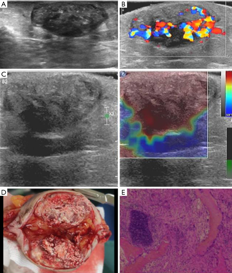
Case description: We report a case of pilomatricoma located on the 47-year-old female breast, characterized by multiple punctate hyperechoic foci internally, presenting as a firm, non-tender mass on palpation and elastography. Without seeking treatment, she experienced pain in the lump two weeks before presentation. After applying an unspecified anti-inflammatory ointment, the lump markedly increased. Therefore, the rapid enlargement of the mass within a short period raised clinical suspicion of breast cancer, leading to surgical excision, which subsequently confirmed pilomatricoma through histopathological examination.
Conclusions: Pilomatricomas occurring on the breast may present clinically and ultrasonographically similar to breast cancer, necessitating a focus on distinguishing between the two entities based on layer-derived differentiation on ultrasound and elastography.

 求助内容:
求助内容: 应助结果提醒方式:


