{"title":"Anatomical variants of the posterior horns of the lateral ventricles: an MRI study.","authors":"Ronen Spierer, Omer Zarrabi Itzhak, Jonathan Gross, Tamer Sobeh, Shai Shrot","doi":"10.3389/fnimg.2025.1478137","DOIUrl":null,"url":null,"abstract":"<p><strong>Introduction: </strong>Anatomical variations in the posterior horns of the lateral ventricles are well-documented, with the horn presenting as open, constricted, or completely closed. However, the extent and nature of these variations across different demographics remain under-explored. This study aimed to investigate the anatomical variations of the posterior horn of the lateral ventricles across different age and sex groups and to compare the variations between the right and left sides.</p><p><strong>Methods: </strong>We conducted a retrospective analysis of magnetic resonance imaging (MRI) scans from 217 adult participants across 15 age groups, utilizing a stratified random sampling from a radiology database. MRI scans were analyzed for ventricular dimensions, and horn types (open, constricted, and closed). Statistical significance was defined as <i>p</i>-value < 0.05.</p><p><strong>Results: </strong>Variants of the posterior horn were observed frequently, with open posterior horn being the most common in the left lateral ventricle (41%) and constricted type being the most common in the right lateral ventricle (37%). A significant correlation existed between the right and left horn types, but in most cases, there was a difference in type between the right and the left horns in the same individual. No significant association between age and the type of the posterior horns was found. However, there was a significant difference in the width and length of the horns between the open and other types, with open horns being wider and longer. Lastly, the left horn appeared longer than the right one.</p><p><strong>Discussion: </strong>The findings underline the high variability in posterior horn morphology, which is not significantly influenced by age or sex but varies between individuals and sides. Future studies should explore the functional impact of these anatomical variations.</p>","PeriodicalId":73094,"journal":{"name":"Frontiers in neuroimaging","volume":"4 ","pages":"1478137"},"PeriodicalIF":0.0000,"publicationDate":"2025-04-07","publicationTypes":"Journal Article","fieldsOfStudy":null,"isOpenAccess":false,"openAccessPdf":"https://www.ncbi.nlm.nih.gov/pmc/articles/PMC12009867/pdf/","citationCount":"0","resultStr":null,"platform":"Semanticscholar","paperid":null,"PeriodicalName":"Frontiers in neuroimaging","FirstCategoryId":"1085","ListUrlMain":"https://doi.org/10.3389/fnimg.2025.1478137","RegionNum":0,"RegionCategory":null,"ArticlePicture":[],"TitleCN":null,"AbstractTextCN":null,"PMCID":null,"EPubDate":"2025/1/1 0:00:00","PubModel":"eCollection","JCR":"","JCRName":"","Score":null,"Total":0}
引用次数: 0
Abstract
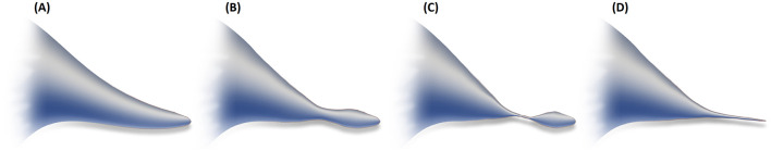
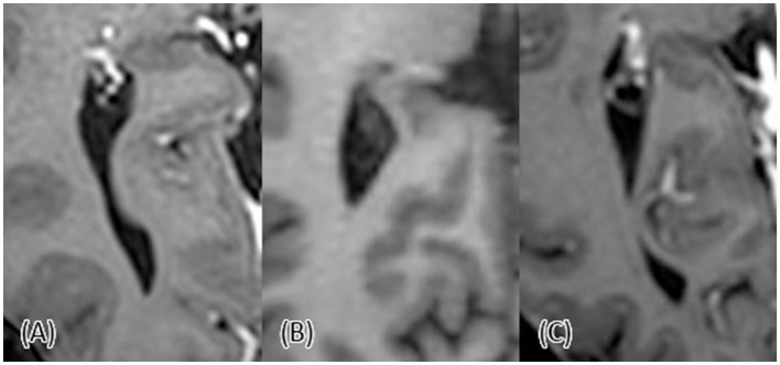
Introduction: Anatomical variations in the posterior horns of the lateral ventricles are well-documented, with the horn presenting as open, constricted, or completely closed. However, the extent and nature of these variations across different demographics remain under-explored. This study aimed to investigate the anatomical variations of the posterior horn of the lateral ventricles across different age and sex groups and to compare the variations between the right and left sides.
Methods: We conducted a retrospective analysis of magnetic resonance imaging (MRI) scans from 217 adult participants across 15 age groups, utilizing a stratified random sampling from a radiology database. MRI scans were analyzed for ventricular dimensions, and horn types (open, constricted, and closed). Statistical significance was defined as p-value < 0.05.
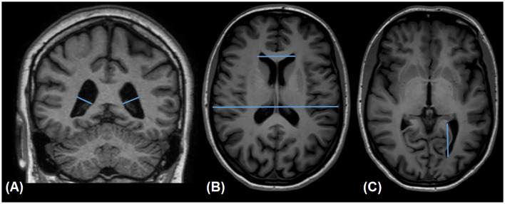
Results: Variants of the posterior horn were observed frequently, with open posterior horn being the most common in the left lateral ventricle (41%) and constricted type being the most common in the right lateral ventricle (37%). A significant correlation existed between the right and left horn types, but in most cases, there was a difference in type between the right and the left horns in the same individual. No significant association between age and the type of the posterior horns was found. However, there was a significant difference in the width and length of the horns between the open and other types, with open horns being wider and longer. Lastly, the left horn appeared longer than the right one.
Discussion: The findings underline the high variability in posterior horn morphology, which is not significantly influenced by age or sex but varies between individuals and sides. Future studies should explore the functional impact of these anatomical variations.

 求助内容:
求助内容: 应助结果提醒方式:


