{"title":"Cytological Diagnosis of Hepatic Metastasis from Rectal Gastrointestinal Stromal Tumor: A Case Report.","authors":"Gauri Niranjan, Pallavi Prasad, Archana Verma, Ashok Kumar","doi":"10.15190/d.2025.1","DOIUrl":null,"url":null,"abstract":"<p><p>Gastrointestinal stromal tumours (GIST) are rare mesenchymal tumours which represent 1% to 3% of all gastrointestinal neoplasms. Rectal location of GIST is extremely rare accounting for 5% of GIST and only 0.1% of rectal tumours. They usually metastasise to the liver (65%). We hereby report a case of rectal stromal tumour with hepatic metastasis. A 55-year-old female presented with pelvic pain, associated with rectal bleeding. A thoracoabdominal computed tomography showed a large heterogeneous enhancing mass, arising from the rectum, anal canal and distal sigmoid colon measuring 12.3x8.7x7.6cm. Based on histopathological examination followed by immunohistochemistry, she was diagnosed with locally advanced rectal GIST. The tumour reduced in size after neoadjuvant-targeted treatment with imatinib. A local resection of the rectal GIST was successfully performed, and a diversion colostomy was done, later colostomy bag was attached. Following the operation, oral imatinib treatment was continued. On subsequent follow-up, her triple phase CECT whole abdomen showed multiple small well-defined peripherally enhancing hypodense liver lesions, the largest measuring 29x18mm suggestive of metastases. Ultrasound-guided fine needle aspiration from a liver lesion was reported as metastatic GIST. The patient underwent surgery, sunitinib was started and was discharged in stable condition. Thus, cytologic examination provides rapid interpretation, is a less invasive technique than open biopsy, and provides a cost-effective modality for diagnosing and managing inaccessible lesions.</p>","PeriodicalId":72829,"journal":{"name":"Discoveries (Craiova, Romania)","volume":"13 1","pages":"e202"},"PeriodicalIF":0.0000,"publicationDate":"2025-03-31","publicationTypes":"Journal Article","fieldsOfStudy":null,"isOpenAccess":false,"openAccessPdf":"https://www.ncbi.nlm.nih.gov/pmc/articles/PMC12062738/pdf/","citationCount":"0","resultStr":null,"platform":"Semanticscholar","paperid":null,"PeriodicalName":"Discoveries (Craiova, Romania)","FirstCategoryId":"1085","ListUrlMain":"https://doi.org/10.15190/d.2025.1","RegionNum":0,"RegionCategory":null,"ArticlePicture":[],"TitleCN":null,"AbstractTextCN":null,"PMCID":null,"EPubDate":"2025/1/1 0:00:00","PubModel":"eCollection","JCR":"","JCRName":"","Score":null,"Total":0}
引用次数: 0
Abstract
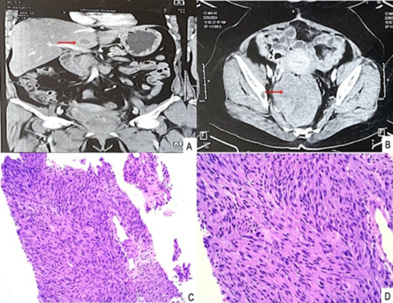
Gastrointestinal stromal tumours (GIST) are rare mesenchymal tumours which represent 1% to 3% of all gastrointestinal neoplasms. Rectal location of GIST is extremely rare accounting for 5% of GIST and only 0.1% of rectal tumours. They usually metastasise to the liver (65%). We hereby report a case of rectal stromal tumour with hepatic metastasis. A 55-year-old female presented with pelvic pain, associated with rectal bleeding. A thoracoabdominal computed tomography showed a large heterogeneous enhancing mass, arising from the rectum, anal canal and distal sigmoid colon measuring 12.3x8.7x7.6cm. Based on histopathological examination followed by immunohistochemistry, she was diagnosed with locally advanced rectal GIST. The tumour reduced in size after neoadjuvant-targeted treatment with imatinib. A local resection of the rectal GIST was successfully performed, and a diversion colostomy was done, later colostomy bag was attached. Following the operation, oral imatinib treatment was continued. On subsequent follow-up, her triple phase CECT whole abdomen showed multiple small well-defined peripherally enhancing hypodense liver lesions, the largest measuring 29x18mm suggestive of metastases. Ultrasound-guided fine needle aspiration from a liver lesion was reported as metastatic GIST. The patient underwent surgery, sunitinib was started and was discharged in stable condition. Thus, cytologic examination provides rapid interpretation, is a less invasive technique than open biopsy, and provides a cost-effective modality for diagnosing and managing inaccessible lesions.

 求助内容:
求助内容: 应助结果提醒方式:


