Kyoung Yoon Lim, Seongbeom Park, Duk L Na, Sang Won Seo, Min Young Chun, Kichang Kwak
{"title":"Quantifying Brain Atrophy Using a CSF-Focused Segmentation Approach.","authors":"Kyoung Yoon Lim, Seongbeom Park, Duk L Na, Sang Won Seo, Min Young Chun, Kichang Kwak","doi":"10.12779/dnd.2025.24.2.115","DOIUrl":null,"url":null,"abstract":"<p><strong>Background and purpose: </strong>Brain atrophy, characterized by sulcal widening and ventricular enlargement, is a hallmark of neurodegenerative diseases such as Alzheimer's disease. Visual assessments are subjective and variable, while automated methods struggle with subtle intensity differences and standardization, highlighting limitations in both approaches. This study aimed to develop and evaluate a novel method focusing on cerebrospinal fluid (CSF) regions by assessing segmentation accuracy, detecting stage-specific atrophy patterns, and testing generalizability to unstandardized datasets.</p><p><strong>Methods: </strong>We utilized T1-weighted magnetic resonance imaging data from 3,315 participants from Samsung Medical Center and 1,439 participants from other hospitals. Segmentation accuracy was evaluated using the Dice similarity coefficient (DSC), and W-scores were calculated for each region of interest (ROI) to assess stage-specific atrophy patterns.</p><p><strong>Results: </strong>The segmentation demonstrated high accuracy, with average DSC values exceeding 0.9 for ventricular and hippocampal regions and above 0.8 for cortical regions. Significant differences in W-scores were observed across cognitive stages (cognitively unimpaired, mild cognitive impairment, dementia of Alzheimer's type) for all ROIs (all, <i>p</i><0.05). Similar trends were observed in the images from other hospitals, confirming the algorithm's generalizability to datasets without prior standardization.</p><p><strong>Conclusions: </strong>This study demonstrates the robustness and clinical applicability of a novel CSF-focused segmentation method for assessing brain atrophy. The method provides a scalable and objective framework for evaluating structural changes across cognitive stages and holds potential for broader application in neurodegenerative disease research and clinical practice.</p>","PeriodicalId":72779,"journal":{"name":"Dementia and neurocognitive disorders","volume":"24 2","pages":"115-125"},"PeriodicalIF":0.0000,"publicationDate":"2025-04-01","publicationTypes":"Journal Article","fieldsOfStudy":null,"isOpenAccess":false,"openAccessPdf":"https://www.ncbi.nlm.nih.gov/pmc/articles/PMC12046248/pdf/","citationCount":"0","resultStr":null,"platform":"Semanticscholar","paperid":null,"PeriodicalName":"Dementia and neurocognitive disorders","FirstCategoryId":"1085","ListUrlMain":"https://doi.org/10.12779/dnd.2025.24.2.115","RegionNum":0,"RegionCategory":null,"ArticlePicture":[],"TitleCN":null,"AbstractTextCN":null,"PMCID":null,"EPubDate":"2025/4/9 0:00:00","PubModel":"Epub","JCR":"","JCRName":"","Score":null,"Total":0}
引用次数: 0
Abstract
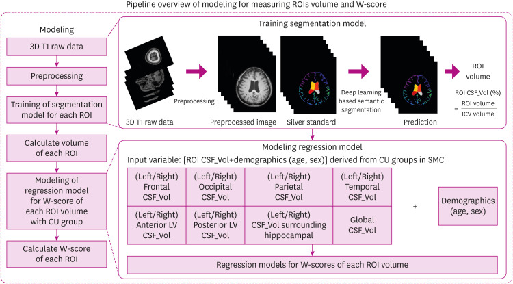
Background and purpose: Brain atrophy, characterized by sulcal widening and ventricular enlargement, is a hallmark of neurodegenerative diseases such as Alzheimer's disease. Visual assessments are subjective and variable, while automated methods struggle with subtle intensity differences and standardization, highlighting limitations in both approaches. This study aimed to develop and evaluate a novel method focusing on cerebrospinal fluid (CSF) regions by assessing segmentation accuracy, detecting stage-specific atrophy patterns, and testing generalizability to unstandardized datasets.
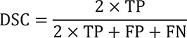
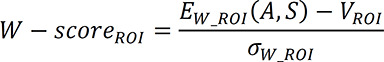
Methods: We utilized T1-weighted magnetic resonance imaging data from 3,315 participants from Samsung Medical Center and 1,439 participants from other hospitals. Segmentation accuracy was evaluated using the Dice similarity coefficient (DSC), and W-scores were calculated for each region of interest (ROI) to assess stage-specific atrophy patterns.
Results: The segmentation demonstrated high accuracy, with average DSC values exceeding 0.9 for ventricular and hippocampal regions and above 0.8 for cortical regions. Significant differences in W-scores were observed across cognitive stages (cognitively unimpaired, mild cognitive impairment, dementia of Alzheimer's type) for all ROIs (all, p<0.05). Similar trends were observed in the images from other hospitals, confirming the algorithm's generalizability to datasets without prior standardization.
Conclusions: This study demonstrates the robustness and clinical applicability of a novel CSF-focused segmentation method for assessing brain atrophy. The method provides a scalable and objective framework for evaluating structural changes across cognitive stages and holds potential for broader application in neurodegenerative disease research and clinical practice.

 求助内容:
求助内容: 应助结果提醒方式:


