{"title":"Changes in the electromyographic activity of masticatory muscles in patients undergoing bimaxillary surgery.","authors":"Ali Kağan Özen, İsmail Ceylan","doi":"10.2340/aos.v84.43408","DOIUrl":null,"url":null,"abstract":"<p><strong>Objective: </strong>The aim of this study is to investigate short-term changes in the electromyographic (EMG) activity of masticatory muscles in individuals with skeletal Class III malocclusion undergo double-jaw orthognathic surgery.</p><p><strong>Material and methods: </strong>In patients with skeletal Class III anomaly, EMG activity changes in the anterior temporalis and masseter muscles were measured before T0 and at 3 (T1) and 6 (T2) months after bimaxillary orthognathic surgery. Recordings were obtained using the 'MP100' device and circular Ag-AgCl electrodes during closure, clenching, chewing and swallowing. Data from 26 individuals (12 males, 14 females) with a mean age of 21.7 years were analysed using the Friedman test.</p><p><strong>Results: </strong>A significant decrease was observed in the right/left masseter muscles during clenching/chewing from T1 to T0 (right and left masseter clenching: p < 0.001, left masseter chewing: p < 0.01, right masseter chewing: p < 0.05), while a significant increase was noted in the right masseter's clenching function from T2 to T1 (p < 0.01). The EMG activity of the right anterior temporal muscle decreased during clenching at T1-T0 (p < 0.001), increased at T2-T1 (p < 0.05), and decreased during chewing/swallowing at T1-T0 (chewing: p < 0.001, swallowing: p < 0.05) and T2-T0 (p < 0.05). The left anterior temporal muscle showed decreased EMG activity during clenching at T1-T0 (p < 0.001), increased at T2-T1 (p < 0.05), and decreased during chewing at T1-T0 (p < 0.001). During swallowing, a decrease was observed at T2-T0 (p < 0.001).</p><p><strong>Conclusions: </strong>Partial changes in EMG activity were observed during some functions in the 3-month period; however, no significant overall change was recorded in the 6-month period.</p>","PeriodicalId":7313,"journal":{"name":"Acta Odontologica Scandinavica","volume":"84 ","pages":"182-190"},"PeriodicalIF":1.9000,"publicationDate":"2025-04-22","publicationTypes":"Journal Article","fieldsOfStudy":null,"isOpenAccess":false,"openAccessPdf":"https://www.ncbi.nlm.nih.gov/pmc/articles/PMC12056318/pdf/","citationCount":"0","resultStr":null,"platform":"Semanticscholar","paperid":null,"PeriodicalName":"Acta Odontologica Scandinavica","FirstCategoryId":"3","ListUrlMain":"https://doi.org/10.2340/aos.v84.43408","RegionNum":4,"RegionCategory":"医学","ArticlePicture":[],"TitleCN":null,"AbstractTextCN":null,"PMCID":null,"EPubDate":"","PubModel":"","JCR":"Q3","JCRName":"DENTISTRY, ORAL SURGERY & MEDICINE","Score":null,"Total":0}
引用次数: 0
Abstract
Objective: The aim of this study is to investigate short-term changes in the electromyographic (EMG) activity of masticatory muscles in individuals with skeletal Class III malocclusion undergo double-jaw orthognathic surgery.
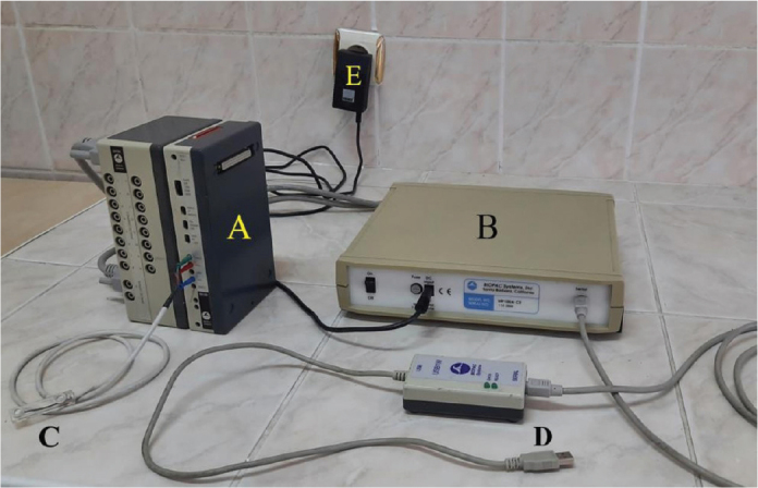
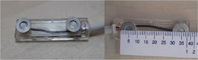
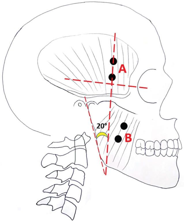
Material and methods: In patients with skeletal Class III anomaly, EMG activity changes in the anterior temporalis and masseter muscles were measured before T0 and at 3 (T1) and 6 (T2) months after bimaxillary orthognathic surgery. Recordings were obtained using the 'MP100' device and circular Ag-AgCl electrodes during closure, clenching, chewing and swallowing. Data from 26 individuals (12 males, 14 females) with a mean age of 21.7 years were analysed using the Friedman test.
Results: A significant decrease was observed in the right/left masseter muscles during clenching/chewing from T1 to T0 (right and left masseter clenching: p < 0.001, left masseter chewing: p < 0.01, right masseter chewing: p < 0.05), while a significant increase was noted in the right masseter's clenching function from T2 to T1 (p < 0.01). The EMG activity of the right anterior temporal muscle decreased during clenching at T1-T0 (p < 0.001), increased at T2-T1 (p < 0.05), and decreased during chewing/swallowing at T1-T0 (chewing: p < 0.001, swallowing: p < 0.05) and T2-T0 (p < 0.05). The left anterior temporal muscle showed decreased EMG activity during clenching at T1-T0 (p < 0.001), increased at T2-T1 (p < 0.05), and decreased during chewing at T1-T0 (p < 0.001). During swallowing, a decrease was observed at T2-T0 (p < 0.001).
Conclusions: Partial changes in EMG activity were observed during some functions in the 3-month period; however, no significant overall change was recorded in the 6-month period.
 求助内容:
求助内容: 应助结果提醒方式:


