Francesco Vasuri, Elisa Albertini, Lucia Miranda, Thais Maloberti, Stefano Chillotti, Sara Coluccelli, Giovanni Tallini, Antonia D'Errico, Dario de Biase
{"title":"Morpho-molecular approach (NGS <i>plus</i> digital PCR) in diagnosis of malignant biliary strictures.","authors":"Francesco Vasuri, Elisa Albertini, Lucia Miranda, Thais Maloberti, Stefano Chillotti, Sara Coluccelli, Giovanni Tallini, Antonia D'Errico, Dario de Biase","doi":"10.32074/1591-951X-1117","DOIUrl":null,"url":null,"abstract":"<p><strong>Objective: </strong>To analyze the diagnostic accuracy and feasibility of <i>digital</i>-PCR (dPCR) combined with next-generation sequencing (NGS) in the ERCP-guided histological diagnosis of biliary strictures to overcome the issue represented by the scarcity of sampled material.</p><p><strong>Methods: </strong>Twenty-two prospective patients were included, and submitted to ERPC-guided biopsy or biliary resection. By histopathological analysis plus fluorescence in situ hybridization (FISH) for chromosomes 3, 7, and 17 aneuploidies, 8 cases (36.4%) were malignant, and 14 cases (63.6%) were negative. NGS was performed on paraffin-embedded tissue by a laboratory-developed panel allowing the analysis of hot-spot regions in 28 genes. Digital PCR (dPCR) was performed by QuantStudio™ AbsoluteQ™ solid dPCR and the copy-number variation (CNV) of the chromosomes 3, 7, and 17 analysed.</p><p><strong>Results: </strong>At dPCR, 1 case showed aneuploidy of chromosome 3, and 2 cases of both chromosomes 3 and 7. These 3 cases all belonged to the positive group (<i>p</i> = 0.014). At NGS, 6 cases showed at least one mutated gene, all in the positive group (<i>p <</i> 0.001). The 3 cases showing aneuploidy at dPCR also showed mutations at NGS. Basing on these observations, we can propose a diagnostic algorithm: dPCR can be applied first, allowing a diagnosis of malignancy in one working day if aneuploidies are observed. In the case of negative dPCR, a \"second-line\" NGS is performed on the same extracted material.</p><p><strong>Conclusions: </strong>The implementation of dPCR allowed the identification of nearly 40% of positive cases in just one working day. In cases of negative dPCR, the NGS procedure can start on the same extracted nucleic acid used for dPCR, requiring more time, but reaching a 75% sensitivity. More studies are required to identify other more sensitive and specific dPCR targets, but even if our algorithm does not increase diagnostic accuracy, the possibility of avoiding FISH and reaching a diagnosis in a more time- and money-saving fashion might be an important step.</p>","PeriodicalId":45893,"journal":{"name":"PATHOLOGICA","volume":"117 1","pages":"10-17"},"PeriodicalIF":2.9000,"publicationDate":"2025-02-01","publicationTypes":"Journal Article","fieldsOfStudy":null,"isOpenAccess":false,"openAccessPdf":"https://www.ncbi.nlm.nih.gov/pmc/articles/PMC11983076/pdf/","citationCount":"0","resultStr":null,"platform":"Semanticscholar","paperid":null,"PeriodicalName":"PATHOLOGICA","FirstCategoryId":"1085","ListUrlMain":"https://doi.org/10.32074/1591-951X-1117","RegionNum":0,"RegionCategory":null,"ArticlePicture":[],"TitleCN":null,"AbstractTextCN":null,"PMCID":null,"EPubDate":"","PubModel":"","JCR":"Q1","JCRName":"PATHOLOGY","Score":null,"Total":0}
引用次数: 0
Abstract
Objective: To analyze the diagnostic accuracy and feasibility of digital-PCR (dPCR) combined with next-generation sequencing (NGS) in the ERCP-guided histological diagnosis of biliary strictures to overcome the issue represented by the scarcity of sampled material.
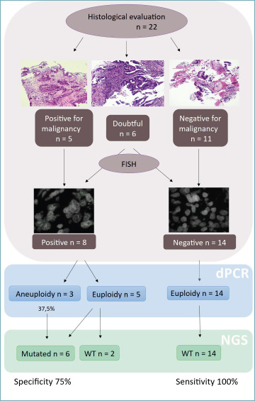
Methods: Twenty-two prospective patients were included, and submitted to ERPC-guided biopsy or biliary resection. By histopathological analysis plus fluorescence in situ hybridization (FISH) for chromosomes 3, 7, and 17 aneuploidies, 8 cases (36.4%) were malignant, and 14 cases (63.6%) were negative. NGS was performed on paraffin-embedded tissue by a laboratory-developed panel allowing the analysis of hot-spot regions in 28 genes. Digital PCR (dPCR) was performed by QuantStudio™ AbsoluteQ™ solid dPCR and the copy-number variation (CNV) of the chromosomes 3, 7, and 17 analysed.
Results: At dPCR, 1 case showed aneuploidy of chromosome 3, and 2 cases of both chromosomes 3 and 7. These 3 cases all belonged to the positive group (p = 0.014). At NGS, 6 cases showed at least one mutated gene, all in the positive group (p < 0.001). The 3 cases showing aneuploidy at dPCR also showed mutations at NGS. Basing on these observations, we can propose a diagnostic algorithm: dPCR can be applied first, allowing a diagnosis of malignancy in one working day if aneuploidies are observed. In the case of negative dPCR, a "second-line" NGS is performed on the same extracted material.
Conclusions: The implementation of dPCR allowed the identification of nearly 40% of positive cases in just one working day. In cases of negative dPCR, the NGS procedure can start on the same extracted nucleic acid used for dPCR, requiring more time, but reaching a 75% sensitivity. More studies are required to identify other more sensitive and specific dPCR targets, but even if our algorithm does not increase diagnostic accuracy, the possibility of avoiding FISH and reaching a diagnosis in a more time- and money-saving fashion might be an important step.

 求助内容:
求助内容: 应助结果提醒方式:


