{"title":"Recalcitrant Fovea-Involving Macular Fold After Uneventful Epiretinal Membrane Surgery.","authors":"Matteo Mario Carlà, Carlos Mateo","doi":"10.22336/rjo.2025.20","DOIUrl":null,"url":null,"abstract":"<p><strong>Purpose: </strong>To describe a case of recalcitrant fovea-involving macular folds developing after uncomplicated epiretinal membrane (ERM) peeling and causing intractable metamorphopsia.</p><p><strong>Methods: </strong>Case report.</p><p><strong>Results: </strong>A 22-year-old man with a stage 3 ERM, visual acuity (VA) of 20/100, and a history of scleral buckle underwent pars plana vitrectomy (PPV) with internal limiting membrane (ILM) peeling. One week after surgery, VA dropped to 20/125 with worsened metamorphopsia. Ophthalmoscopy revealed a fovea-involving full-thickness macular fold, with photoreceptor outer segments in opposition. After 3 weeks of follow-up without improvement, PPV with induction of localized retinal detachment was performed, combined with retinal massage. Moreover, perfluorocarbon liquid (PFCL) was employed to stretch the retina. Nevertheless, the macular fold and metamorphopsia were unchanged, even one year after the first surgery.</p><p><strong>Discussion: </strong>We hypothesize that, due to the highly contracted ERM, the retina may have separated from the RPE during peeling and folded over in the first postoperative days. Concurrently, incorrect patient positioning under air tamponade might have contributed to the vertical orientation of the fold.</p><p><strong>Conclusion: </strong>Even if macular folds after ERM surgery are rare, prompt surgical treatment rather than watchful waiting should be considered to prevent permanent functional impairment.</p>","PeriodicalId":94355,"journal":{"name":"Romanian journal of ophthalmology","volume":"69 1","pages":"124-128"},"PeriodicalIF":0.0000,"publicationDate":"2025-01-01","publicationTypes":"Journal Article","fieldsOfStudy":null,"isOpenAccess":false,"openAccessPdf":"https://www.ncbi.nlm.nih.gov/pmc/articles/PMC12049659/pdf/","citationCount":"0","resultStr":null,"platform":"Semanticscholar","paperid":null,"PeriodicalName":"Romanian journal of ophthalmology","FirstCategoryId":"1085","ListUrlMain":"https://doi.org/10.22336/rjo.2025.20","RegionNum":0,"RegionCategory":null,"ArticlePicture":[],"TitleCN":null,"AbstractTextCN":null,"PMCID":null,"EPubDate":"","PubModel":"","JCR":"","JCRName":"","Score":null,"Total":0}
引用次数: 0
Abstract
Purpose: To describe a case of recalcitrant fovea-involving macular folds developing after uncomplicated epiretinal membrane (ERM) peeling and causing intractable metamorphopsia.
Methods: Case report.
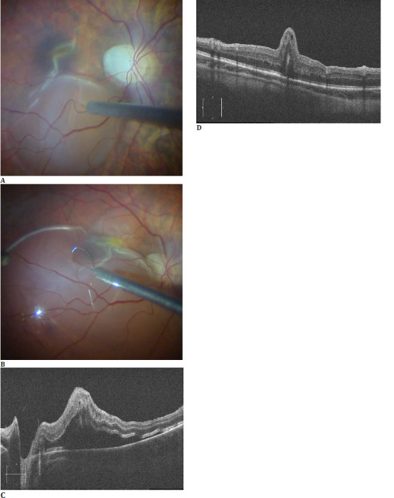
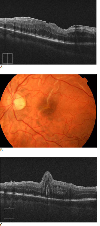
Results: A 22-year-old man with a stage 3 ERM, visual acuity (VA) of 20/100, and a history of scleral buckle underwent pars plana vitrectomy (PPV) with internal limiting membrane (ILM) peeling. One week after surgery, VA dropped to 20/125 with worsened metamorphopsia. Ophthalmoscopy revealed a fovea-involving full-thickness macular fold, with photoreceptor outer segments in opposition. After 3 weeks of follow-up without improvement, PPV with induction of localized retinal detachment was performed, combined with retinal massage. Moreover, perfluorocarbon liquid (PFCL) was employed to stretch the retina. Nevertheless, the macular fold and metamorphopsia were unchanged, even one year after the first surgery.
Discussion: We hypothesize that, due to the highly contracted ERM, the retina may have separated from the RPE during peeling and folded over in the first postoperative days. Concurrently, incorrect patient positioning under air tamponade might have contributed to the vertical orientation of the fold.
Conclusion: Even if macular folds after ERM surgery are rare, prompt surgical treatment rather than watchful waiting should be considered to prevent permanent functional impairment.

 求助内容:
求助内容: 应助结果提醒方式:


