{"title":"Bilateral Optic Atrophy and Epiretinal Membranes: An Atypical Presentation of Ocular Tuberculosis.","authors":"Yewande Olubunmi Babalola","doi":"10.71480/nmj.v66i1.711","DOIUrl":null,"url":null,"abstract":"<p><p>A 15-year-old boy presented himself to the retina clinic with a six-year history of poor vision in both eyes. Ocular symptoms started with deterioration in the vision of both eyes associated with ocular pain. There was no history of antecedent trauma, headaches, seizures, or loss of consciousness. The general and systemic examinations were normal. On ocular examination, the corrected visual acuity was 6/24 and 6/6 in the right and left eyes, respectively. The anterior segment examination was normal except for a relative afferent pupillary defect in the right eye. Examination of the posterior segment revealed bilateral optic atrophy worse in the right eye and cup disc ratios of 0.3 bilaterally. The vessels were within normal limits with epiretinal membranes in both eyes. Yellowish chorioretinal lesions were present temporal to the disc in both eyes. There was a positive history of tuberculosis in the father and brother. A diagnosis of bilateral optic atrophy and epiretinal membranes secondary to presumed ocular tuberculosis was made.</p>","PeriodicalId":94346,"journal":{"name":"Nigerian medical journal : journal of the Nigeria Medical Association","volume":"66 1","pages":"389-393"},"PeriodicalIF":0.0000,"publicationDate":"2025-04-03","publicationTypes":"Journal Article","fieldsOfStudy":null,"isOpenAccess":false,"openAccessPdf":"https://www.ncbi.nlm.nih.gov/pmc/articles/PMC12038614/pdf/","citationCount":"0","resultStr":null,"platform":"Semanticscholar","paperid":null,"PeriodicalName":"Nigerian medical journal : journal of the Nigeria Medical Association","FirstCategoryId":"1085","ListUrlMain":"https://doi.org/10.71480/nmj.v66i1.711","RegionNum":0,"RegionCategory":null,"ArticlePicture":[],"TitleCN":null,"AbstractTextCN":null,"PMCID":null,"EPubDate":"2025/1/1 0:00:00","PubModel":"eCollection","JCR":"","JCRName":"","Score":null,"Total":0}
引用次数: 0
Abstract
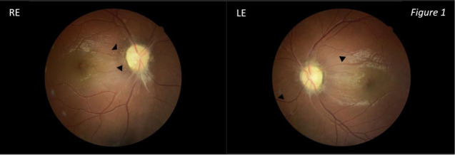
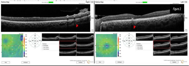
A 15-year-old boy presented himself to the retina clinic with a six-year history of poor vision in both eyes. Ocular symptoms started with deterioration in the vision of both eyes associated with ocular pain. There was no history of antecedent trauma, headaches, seizures, or loss of consciousness. The general and systemic examinations were normal. On ocular examination, the corrected visual acuity was 6/24 and 6/6 in the right and left eyes, respectively. The anterior segment examination was normal except for a relative afferent pupillary defect in the right eye. Examination of the posterior segment revealed bilateral optic atrophy worse in the right eye and cup disc ratios of 0.3 bilaterally. The vessels were within normal limits with epiretinal membranes in both eyes. Yellowish chorioretinal lesions were present temporal to the disc in both eyes. There was a positive history of tuberculosis in the father and brother. A diagnosis of bilateral optic atrophy and epiretinal membranes secondary to presumed ocular tuberculosis was made.

 求助内容:
求助内容: 应助结果提醒方式:


