Meihai Xu, Zheng Wang, Xiu-Feng Qiao, Hai Liao, Dan-Ke Su
{"title":"A nomogram model for predicting lymph node metastasis of rectal cancer by combining preoperative magnetic resonance imaging signs and tumour markers.","authors":"Meihai Xu, Zheng Wang, Xiu-Feng Qiao, Hai Liao, Dan-Ke Su","doi":"10.5114/pjr/200612","DOIUrl":null,"url":null,"abstract":"<p><strong>Purpose: </strong>This study aimed to explore the diagnostic value of high-resolution magnetic resonance images and tumour markers in predicting lymph node metastasis of rectal cancer.</p><p><strong>Material and methods: </strong>The clinical, imaging, and pathological data of patients with suspected rectal cancer were collected. The baseline data, and surgical and pathological characteristics were compared between the lymph node metastasis group and no metastasis group. Univariate and multivariate logistic regression were used to analyse the clinical and pathological factors, and preoperative magnetic resonance imaging (MRI) signs of extramural vascular invasion and rectal cancer lymph node metastasis. A nomogram model was established with statistically significant factors.</p><p><strong>Results: </strong>150 patients were included. Among them, 50 (33.3%) presented with vascular tumour thrombus, and 72 (48.0%) had lymph node metastasis. The detection of regional lymph nodes (DWI-LN) was an independent risk factor for lymph node metastasis. The area under curve of the nomogram model was 0.804.</p><p><strong>Conclusion: </strong>Preoperative serum CA19.9, and the relationship between tumour and peritoneal reflection in preoperative MRI and DWI-LN have clinical value in predicting lymph node metastasis in patients with rectal cancer.</p>","PeriodicalId":94174,"journal":{"name":"Polish journal of radiology","volume":"90 ","pages":"e114-e123"},"PeriodicalIF":0.0000,"publicationDate":"2025-03-07","publicationTypes":"Journal Article","fieldsOfStudy":null,"isOpenAccess":false,"openAccessPdf":"https://www.ncbi.nlm.nih.gov/pmc/articles/PMC12049155/pdf/","citationCount":"0","resultStr":null,"platform":"Semanticscholar","paperid":null,"PeriodicalName":"Polish journal of radiology","FirstCategoryId":"1085","ListUrlMain":"https://doi.org/10.5114/pjr/200612","RegionNum":0,"RegionCategory":null,"ArticlePicture":[],"TitleCN":null,"AbstractTextCN":null,"PMCID":null,"EPubDate":"2025/1/1 0:00:00","PubModel":"eCollection","JCR":"","JCRName":"","Score":null,"Total":0}
引用次数: 0
Abstract
Purpose: This study aimed to explore the diagnostic value of high-resolution magnetic resonance images and tumour markers in predicting lymph node metastasis of rectal cancer.
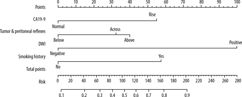
Material and methods: The clinical, imaging, and pathological data of patients with suspected rectal cancer were collected. The baseline data, and surgical and pathological characteristics were compared between the lymph node metastasis group and no metastasis group. Univariate and multivariate logistic regression were used to analyse the clinical and pathological factors, and preoperative magnetic resonance imaging (MRI) signs of extramural vascular invasion and rectal cancer lymph node metastasis. A nomogram model was established with statistically significant factors.
Results: 150 patients were included. Among them, 50 (33.3%) presented with vascular tumour thrombus, and 72 (48.0%) had lymph node metastasis. The detection of regional lymph nodes (DWI-LN) was an independent risk factor for lymph node metastasis. The area under curve of the nomogram model was 0.804.
Conclusion: Preoperative serum CA19.9, and the relationship between tumour and peritoneal reflection in preoperative MRI and DWI-LN have clinical value in predicting lymph node metastasis in patients with rectal cancer.

 求助内容:
求助内容: 应助结果提醒方式:


