Andric Pérez-Ortiz, Ana Yolanda Sandoval-Mussi, Anallely Luna-Hernández, Antonio Giovanni Spaventa-Ibarrola, Antonio Herrera-González, César Manuel Vargas-Sahagun, Carlos Zerrweck
{"title":"[Laparoscopic management of gastric schwannoma].","authors":"Andric Pérez-Ortiz, Ana Yolanda Sandoval-Mussi, Anallely Luna-Hernández, Antonio Giovanni Spaventa-Ibarrola, Antonio Herrera-González, César Manuel Vargas-Sahagun, Carlos Zerrweck","doi":"10.5281/zenodo.14617155","DOIUrl":null,"url":null,"abstract":"<p><strong>Background: </strong>Gastric schwannoma is a rare GI tract tumor. Commonly, cases are asymptomatic and often misdiagnosed as gastrointestinal stromal tumors (GIST) or gastric leiomyomas.</p><p><strong>Clinical case: </strong>A 34-year-old female presented to the clinic for a gastric sleeve for obesity. Preoperatively, there was a GIST misdiagnosis. A CT scan showed a 16 mm gastric tumor in the lesser curvature adjacent to segment III of the liver. Endoscopic ultrasound showed a lesion arising from the muscularis mucosa. The biopsy was positive for spindle cells. The patient underwent a laparoscopic resection and Roux-en-Y gastric bypass. The pathology report showed S100-positive spindle cells compatible with gastric schwannoma.</p><p><strong>Conclusions: </strong>Like our case, gastric schwannomas are usually asymptomatic and arise in middle-aged women. However, their submucosal location in imaging studies, even endoscopic ultrasound, often leads to misdiagnosis. Postoperative pathology and immunohistochemical examinations are critical for final diagnosis and distinguishing features of other malignant stromal cell tumors. Care must be taken in cases with prominent lymph nodes on diagnostic laparoscopy.</p>","PeriodicalId":94200,"journal":{"name":"Revista medica del Instituto Mexicano del Seguro Social","volume":"63 2","pages":"e6338"},"PeriodicalIF":0.0000,"publicationDate":"2025-03-03","publicationTypes":"Journal Article","fieldsOfStudy":null,"isOpenAccess":false,"openAccessPdf":"https://www.ncbi.nlm.nih.gov/pmc/articles/PMC12052381/pdf/","citationCount":"0","resultStr":null,"platform":"Semanticscholar","paperid":null,"PeriodicalName":"Revista medica del Instituto Mexicano del Seguro Social","FirstCategoryId":"1085","ListUrlMain":"https://doi.org/10.5281/zenodo.14617155","RegionNum":0,"RegionCategory":null,"ArticlePicture":[],"TitleCN":null,"AbstractTextCN":null,"PMCID":null,"EPubDate":"","PubModel":"","JCR":"","JCRName":"","Score":null,"Total":0}
引用次数: 0
Abstract
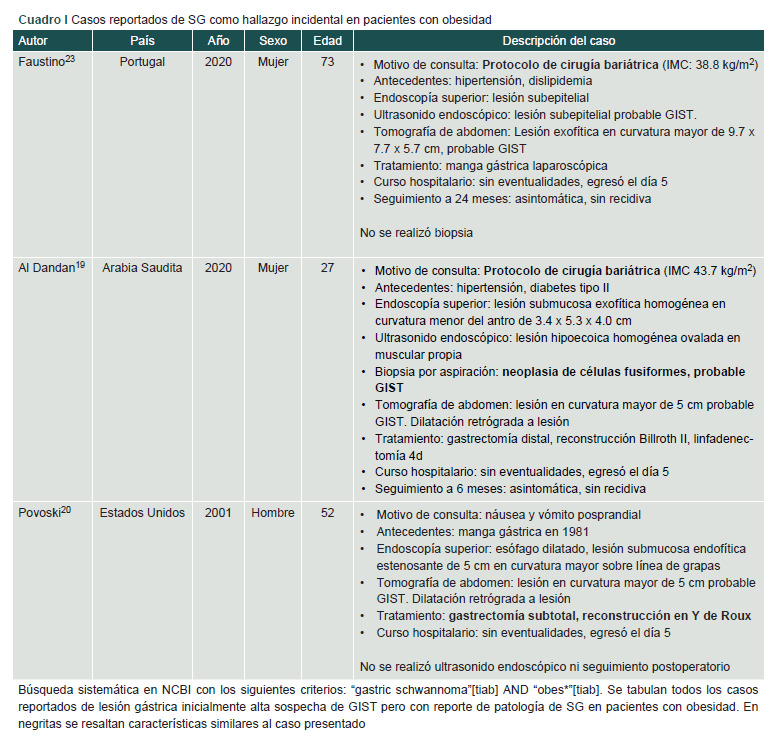
Background: Gastric schwannoma is a rare GI tract tumor. Commonly, cases are asymptomatic and often misdiagnosed as gastrointestinal stromal tumors (GIST) or gastric leiomyomas.
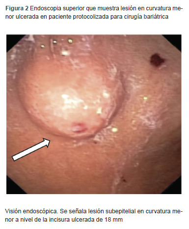
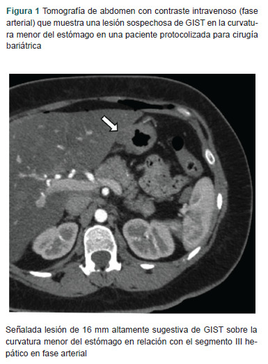
Clinical case: A 34-year-old female presented to the clinic for a gastric sleeve for obesity. Preoperatively, there was a GIST misdiagnosis. A CT scan showed a 16 mm gastric tumor in the lesser curvature adjacent to segment III of the liver. Endoscopic ultrasound showed a lesion arising from the muscularis mucosa. The biopsy was positive for spindle cells. The patient underwent a laparoscopic resection and Roux-en-Y gastric bypass. The pathology report showed S100-positive spindle cells compatible with gastric schwannoma.
Conclusions: Like our case, gastric schwannomas are usually asymptomatic and arise in middle-aged women. However, their submucosal location in imaging studies, even endoscopic ultrasound, often leads to misdiagnosis. Postoperative pathology and immunohistochemical examinations are critical for final diagnosis and distinguishing features of other malignant stromal cell tumors. Care must be taken in cases with prominent lymph nodes on diagnostic laparoscopy.

 求助内容:
求助内容: 应助结果提醒方式:


