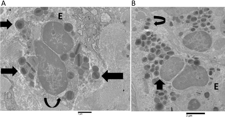
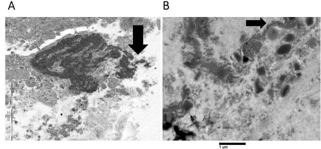
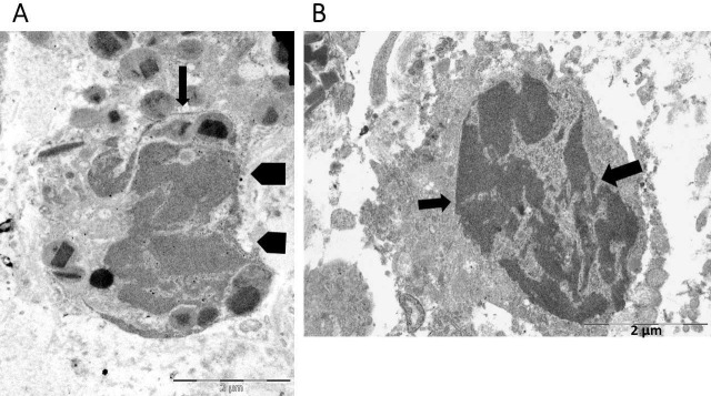
{"title":"Eosinophil cytolysis with or without ETosis in four cases of human gastric cancer: a comparative ultrastructural study.","authors":"Rosario Caruso, Valerio Caruso, Luciana Rigoli","doi":"10.37349/etat.2025.1002309","DOIUrl":null,"url":null,"abstract":"<p><p>The ultrastructural morphology of eosinophil cytolysis and extracellular trap cell death (ETosis) has predominantly been examined in non-neoplastic eosinophil-associated diseases, with a limited investigation in neoplasms. This current electron microscopy study examined the ultrastructural characteristics of eosinophil cytolysis and ETosis across four distinct gastric cancer cases: three cases (cases 1-3) exhibited non-ETotic cytolysis, while one case (case 4) presented eosinophils at various stages of ETosis. In cases 1-3, eosinophil non-ETotic cytolysis was characterized by localized plasma membrane disruption, the presence of free extracellular granules (FEGs), and the maintenance of a round or oval nuclear lobe profile. In case 4, eosinophils were observed in progressive stages of ETosis, arbitrarily subdivided into early, intermediate, and advanced. Although early ETosis and non-ETotic cytolysis exhibited overlapping ultrastructural features, chromatin decondensation and nuclear envelope enlargement were more pronounced in early ETosis. Nuclear envelope disruption, loss of the round or oval nuclear lobe profile (intermediate stage), extracellular DNA trap deposition, and the appearance of Charcot-Leyden crystals (advanced stage) were all distinctive features of ETosis. The findings of this case report confirm previous observations of eosinophil cytolysis with or without ETosis in non-neoplastic diseases and extend them to advanced gastric carcinoma. Since Charcot-Leyden crystals were only seen in case 4, their correlation with ETosis was further supported. In gastric cancer, the release of FEGs during non-ETotic cytolysis and the release of both FEGs and DNA traps during ETotic cytolysis may contribute to the formation of an antitumor microenvironment.</p>","PeriodicalId":73002,"journal":{"name":"Exploration of targeted anti-tumor therapy","volume":"6 ","pages":"1002309"},"PeriodicalIF":0.0000,"publicationDate":"2025-04-21","publicationTypes":"Journal Article","fieldsOfStudy":null,"isOpenAccess":false,"openAccessPdf":"https://www.ncbi.nlm.nih.gov/pmc/articles/PMC12022494/pdf/","citationCount":"0","resultStr":null,"platform":"Semanticscholar","paperid":null,"PeriodicalName":"Exploration of targeted anti-tumor therapy","FirstCategoryId":"1085","ListUrlMain":"https://doi.org/10.37349/etat.2025.1002309","RegionNum":0,"RegionCategory":null,"ArticlePicture":[],"TitleCN":null,"AbstractTextCN":null,"PMCID":null,"EPubDate":"2025/1/1 0:00:00","PubModel":"eCollection","JCR":"Q3","JCRName":"Medicine","Score":null,"Total":0}
引用次数: 0
Abstract
The ultrastructural morphology of eosinophil cytolysis and extracellular trap cell death (ETosis) has predominantly been examined in non-neoplastic eosinophil-associated diseases, with a limited investigation in neoplasms. This current electron microscopy study examined the ultrastructural characteristics of eosinophil cytolysis and ETosis across four distinct gastric cancer cases: three cases (cases 1-3) exhibited non-ETotic cytolysis, while one case (case 4) presented eosinophils at various stages of ETosis. In cases 1-3, eosinophil non-ETotic cytolysis was characterized by localized plasma membrane disruption, the presence of free extracellular granules (FEGs), and the maintenance of a round or oval nuclear lobe profile. In case 4, eosinophils were observed in progressive stages of ETosis, arbitrarily subdivided into early, intermediate, and advanced. Although early ETosis and non-ETotic cytolysis exhibited overlapping ultrastructural features, chromatin decondensation and nuclear envelope enlargement were more pronounced in early ETosis. Nuclear envelope disruption, loss of the round or oval nuclear lobe profile (intermediate stage), extracellular DNA trap deposition, and the appearance of Charcot-Leyden crystals (advanced stage) were all distinctive features of ETosis. The findings of this case report confirm previous observations of eosinophil cytolysis with or without ETosis in non-neoplastic diseases and extend them to advanced gastric carcinoma. Since Charcot-Leyden crystals were only seen in case 4, their correlation with ETosis was further supported. In gastric cancer, the release of FEGs during non-ETotic cytolysis and the release of both FEGs and DNA traps during ETotic cytolysis may contribute to the formation of an antitumor microenvironment.

 求助内容:
求助内容: 应助结果提醒方式:


