{"title":"Comparisons of turbo spin-echo and echo-planar diffusion-weighted imaging of the female pelvic region on 1.5T and 3.0T magnetic resonance systems.","authors":"Hisatoshi Araki, Takeshi Yoshizako, Rika Yoshida, Hiroya Asou, Yasushi Kaji","doi":"10.1177/20584601251339018","DOIUrl":null,"url":null,"abstract":"<p><strong>Background: </strong>There have been no previous studies comparing the effectiveness of echo-planar imaging (EPI)-diffusion-weighted imaging (DWI) and turbo spin echo (TSE)-DWI for female pelvic imaging on 1.5T and 3.0T magnetic resonance imaging (MRI) systems.</p><p><strong>Purpose: </strong>This retrospective study compared EPI-DWI and TSE-DWI of the female pelvis at 1.5T and 3.0T MRI to evaluate image quality, geometric distortion, and apparent diffusion coefficient (ADC) values for optimal clinical diagnosis.</p><p><strong>Materials and methods: </strong>Thirty-nine patients at 1.5T and 71 patients at 3.0T underwent both TSE- and EPI-DWI with b-values of 0 and 1000 s/mm<sup>2</sup>. Spatial resolution was matched between systems and sequences. Geometric distortion, contrast ((uterine myometrium signal intensity (SI) - gluteal muscle SI)/(uterine myometrium SI + gluteal muscles SI)), and ADC values of the uterine myometrium and lesions were compared. Qualitative assessment included ghosting artifacts, image contrast, and overall image quality.</p><p><strong>Results: </strong>There was no significant difference in patient demographics between 1.5T and 3.0T groups. TSE-DWI demonstrated significantly less distortion than EPI-DWI at both field strengths (<i>p</i> < .05). Contrast was higher with TSE-DWI than EPI-DWI at 3.0T (<i>p</i> < .05) and was greater at 3.0T than 1.5T. ADC values of malignancies differed significantly from the uterus and benign lesions across all sequences and field strengths (<i>p</i> < .05). TSE-DWI showed higher ghosting artifact scores than EPI-DWI at 3.0T (<i>p</i> = .019), but no other significant differences in qualitative evaluation were noted.</p><p><strong>Conclusion: </strong>TSE-DWI provided superior contrast with reduced distortion compared to EPI-DWI in the female pelvis at both field strengths, with TSE-DWI demonstrating greater effectiveness at 3.0T.</p>","PeriodicalId":72063,"journal":{"name":"Acta radiologica open","volume":"14 5","pages":"20584601251339018"},"PeriodicalIF":1.0000,"publicationDate":"2025-05-02","publicationTypes":"Journal Article","fieldsOfStudy":null,"isOpenAccess":false,"openAccessPdf":"https://www.ncbi.nlm.nih.gov/pmc/articles/PMC12049636/pdf/","citationCount":"0","resultStr":null,"platform":"Semanticscholar","paperid":null,"PeriodicalName":"Acta radiologica open","FirstCategoryId":"1085","ListUrlMain":"https://doi.org/10.1177/20584601251339018","RegionNum":0,"RegionCategory":null,"ArticlePicture":[],"TitleCN":null,"AbstractTextCN":null,"PMCID":null,"EPubDate":"2025/5/1 0:00:00","PubModel":"eCollection","JCR":"Q4","JCRName":"RADIOLOGY, NUCLEAR MEDICINE & MEDICAL IMAGING","Score":null,"Total":0}
引用次数: 0
Abstract
Background: There have been no previous studies comparing the effectiveness of echo-planar imaging (EPI)-diffusion-weighted imaging (DWI) and turbo spin echo (TSE)-DWI for female pelvic imaging on 1.5T and 3.0T magnetic resonance imaging (MRI) systems.
Purpose: This retrospective study compared EPI-DWI and TSE-DWI of the female pelvis at 1.5T and 3.0T MRI to evaluate image quality, geometric distortion, and apparent diffusion coefficient (ADC) values for optimal clinical diagnosis.
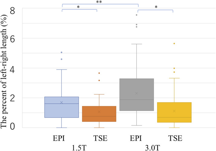
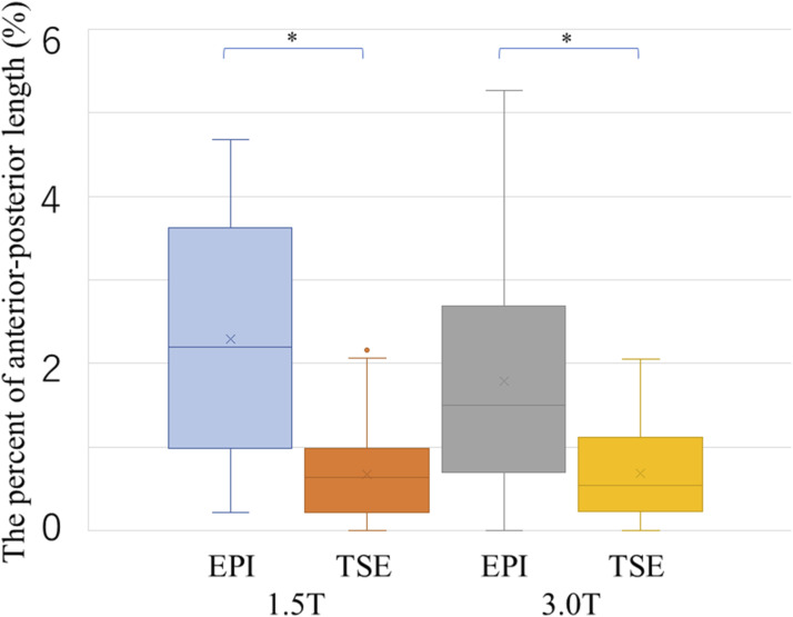
Materials and methods: Thirty-nine patients at 1.5T and 71 patients at 3.0T underwent both TSE- and EPI-DWI with b-values of 0 and 1000 s/mm2. Spatial resolution was matched between systems and sequences. Geometric distortion, contrast ((uterine myometrium signal intensity (SI) - gluteal muscle SI)/(uterine myometrium SI + gluteal muscles SI)), and ADC values of the uterine myometrium and lesions were compared. Qualitative assessment included ghosting artifacts, image contrast, and overall image quality.
Results: There was no significant difference in patient demographics between 1.5T and 3.0T groups. TSE-DWI demonstrated significantly less distortion than EPI-DWI at both field strengths (p < .05). Contrast was higher with TSE-DWI than EPI-DWI at 3.0T (p < .05) and was greater at 3.0T than 1.5T. ADC values of malignancies differed significantly from the uterus and benign lesions across all sequences and field strengths (p < .05). TSE-DWI showed higher ghosting artifact scores than EPI-DWI at 3.0T (p = .019), but no other significant differences in qualitative evaluation were noted.
Conclusion: TSE-DWI provided superior contrast with reduced distortion compared to EPI-DWI in the female pelvis at both field strengths, with TSE-DWI demonstrating greater effectiveness at 3.0T.


 求助内容:
求助内容: 应助结果提醒方式:


