Cynthia Lopes Pereira de Borborema, Eduardo Oliveira Pacheco, Aley Talans, Lucas Rios Torres, Angela Hissae Motoyama Caiado, Felipe Lazar Junior, Ulysses Dos Santos Torres, Giuseppe D'Ippolito
{"title":"Hydatidiform Mole and coexisting fetus following intrauterine insemination: a case report.","authors":"Cynthia Lopes Pereira de Borborema, Eduardo Oliveira Pacheco, Aley Talans, Lucas Rios Torres, Angela Hissae Motoyama Caiado, Felipe Lazar Junior, Ulysses Dos Santos Torres, Giuseppe D'Ippolito","doi":"10.5935/1518-0557.20250011","DOIUrl":null,"url":null,"abstract":"<p><p>Gestational trophoblastic diseases (GTD) comprise a heterogeneous group of disorders arising from genetic anomalies occurring during fertilization in twin pregnancies and often may be associated with assisted reproductive techniques. An exceedingly rare presentation of GTD is a twin pregnancy hydatidiform mole with a co-existing fetus, condition which may be an important cause of complications for the mother and the fetus. A 36-year-old woman (G2, P0, A1) underwent a friendly controlled ovarian stimulation (COS) followed by intrauterine insemination (IUI) for assisted reproductive purposes, resulting in a twin pregnancy initially characterized by two gestational sacs. However, one sac failed to progress and instead degenerated into molar trophoblastic disease, while the other sustained a normal fetus with regular growth. At 33 weeks gestation, the patient developed preeclampsia, necessitating delivery via cesarean section at a tertiary care facility. Reproductive-assisted procedures may be linked to cases of trophoblastic disease. Additionally, the presence of cystic lesions warrants a wide differential diagnosis, with magnetic resonance imaging serving as a valuable tool for accurate assessment and differentiation of structures.</p>","PeriodicalId":46364,"journal":{"name":"Jornal Brasileiro de Reproducao Assistida","volume":" ","pages":"403-406"},"PeriodicalIF":1.9000,"publicationDate":"2025-07-30","publicationTypes":"Journal Article","fieldsOfStudy":null,"isOpenAccess":false,"openAccessPdf":"https://www.ncbi.nlm.nih.gov/pmc/articles/PMC12225151/pdf/","citationCount":"0","resultStr":null,"platform":"Semanticscholar","paperid":null,"PeriodicalName":"Jornal Brasileiro de Reproducao Assistida","FirstCategoryId":"1085","ListUrlMain":"https://doi.org/10.5935/1518-0557.20250011","RegionNum":0,"RegionCategory":null,"ArticlePicture":[],"TitleCN":null,"AbstractTextCN":null,"PMCID":null,"EPubDate":"","PubModel":"","JCR":"Q3","JCRName":"OBSTETRICS & GYNECOLOGY","Score":null,"Total":0}
引用次数: 0
Abstract
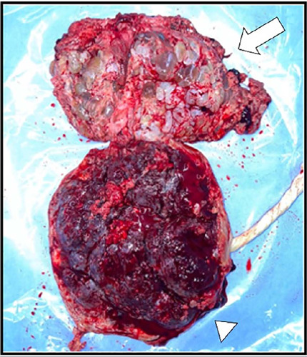
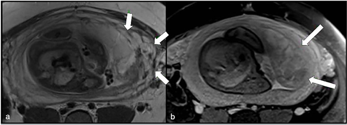
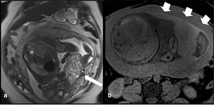
Gestational trophoblastic diseases (GTD) comprise a heterogeneous group of disorders arising from genetic anomalies occurring during fertilization in twin pregnancies and often may be associated with assisted reproductive techniques. An exceedingly rare presentation of GTD is a twin pregnancy hydatidiform mole with a co-existing fetus, condition which may be an important cause of complications for the mother and the fetus. A 36-year-old woman (G2, P0, A1) underwent a friendly controlled ovarian stimulation (COS) followed by intrauterine insemination (IUI) for assisted reproductive purposes, resulting in a twin pregnancy initially characterized by two gestational sacs. However, one sac failed to progress and instead degenerated into molar trophoblastic disease, while the other sustained a normal fetus with regular growth. At 33 weeks gestation, the patient developed preeclampsia, necessitating delivery via cesarean section at a tertiary care facility. Reproductive-assisted procedures may be linked to cases of trophoblastic disease. Additionally, the presence of cystic lesions warrants a wide differential diagnosis, with magnetic resonance imaging serving as a valuable tool for accurate assessment and differentiation of structures.

 求助内容:
求助内容: 应助结果提醒方式:


