{"title":"The ability of plain radiography to accurately describe the bone surface at the head-neck junction of the femur: a study using human bone models.","authors":"Tomohiro Mimura, Yuki Furuya, Kosuke Kumagai, Yasutaka Amano, Shunichi Miyahara, Ryota Uemura, Sadafumi Horikawa, Hideki Saito, Kohei Umeda, Fumitaka Ushiyama, Yugen Ogata, Takafumi Yayama, Kanji Mori, Shinji Imai","doi":"10.1093/jhps/hnae048","DOIUrl":null,"url":null,"abstract":"<p><p>In evaluations of a cam deformity on femoroacetabular impingement, the head-neck junction (HNJ) must be accurately assessed. We conducted this study to determine the ability of plain radiography to visualize the end-to-end bone surface of the HNJ. We used six human bone models. Ten examiners evaluated the degree to which attached stainless wire marker at the 1:00, 1:30, and 2:00 radial plane defined in reconstructed computed tomography can be accurately detected on the bone surface on plain radiographies. We employed 13 plain radiographies: the cross-table lateral view, frog-leg lateral view, Espié frog-leg lateral view, false-profile view, modified false-profile view, 30° Dunn view (DV), 45° DV, 60° DV, 90° DV, 30° modified Dunn view (MDV), 45° MDV, 60° MDV, and 90° MDV. Examiners scored the degree to which the radiographic images accurately detected the stainless wire marker on the bone surface of the HNJ on a scale of 1 point (0% match) to 5 points (almost 100% match). The highest score for the 1:00 plane was 4.98 points on the 45° DV. Similarly, the highest scores of the 1:30 and 2:00 planes were 4.98 points for the 45° MDV and 4.68 points for the 90° MDV, respectively. On these bone model studies, the most suitable plain radiography for describing the HNJ at the 1:00, 1:30, and 2:00 planes were both the 45° DV, the 45° MDV, and the 90° MDV, respectively.</p>","PeriodicalId":48583,"journal":{"name":"Journal of Hip Preservation Surgery","volume":"12 1","pages":"65-73"},"PeriodicalIF":1.1000,"publicationDate":"2024-12-25","publicationTypes":"Journal Article","fieldsOfStudy":null,"isOpenAccess":false,"openAccessPdf":"https://www.ncbi.nlm.nih.gov/pmc/articles/PMC12051853/pdf/","citationCount":"0","resultStr":null,"platform":"Semanticscholar","paperid":null,"PeriodicalName":"Journal of Hip Preservation Surgery","FirstCategoryId":"3","ListUrlMain":"https://doi.org/10.1093/jhps/hnae048","RegionNum":4,"RegionCategory":"医学","ArticlePicture":[],"TitleCN":null,"AbstractTextCN":null,"PMCID":null,"EPubDate":"2025/1/1 0:00:00","PubModel":"eCollection","JCR":"Q3","JCRName":"ORTHOPEDICS","Score":null,"Total":0}
引用次数: 0
Abstract
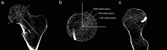
In evaluations of a cam deformity on femoroacetabular impingement, the head-neck junction (HNJ) must be accurately assessed. We conducted this study to determine the ability of plain radiography to visualize the end-to-end bone surface of the HNJ. We used six human bone models. Ten examiners evaluated the degree to which attached stainless wire marker at the 1:00, 1:30, and 2:00 radial plane defined in reconstructed computed tomography can be accurately detected on the bone surface on plain radiographies. We employed 13 plain radiographies: the cross-table lateral view, frog-leg lateral view, Espié frog-leg lateral view, false-profile view, modified false-profile view, 30° Dunn view (DV), 45° DV, 60° DV, 90° DV, 30° modified Dunn view (MDV), 45° MDV, 60° MDV, and 90° MDV. Examiners scored the degree to which the radiographic images accurately detected the stainless wire marker on the bone surface of the HNJ on a scale of 1 point (0% match) to 5 points (almost 100% match). The highest score for the 1:00 plane was 4.98 points on the 45° DV. Similarly, the highest scores of the 1:30 and 2:00 planes were 4.98 points for the 45° MDV and 4.68 points for the 90° MDV, respectively. On these bone model studies, the most suitable plain radiography for describing the HNJ at the 1:00, 1:30, and 2:00 planes were both the 45° DV, the 45° MDV, and the 90° MDV, respectively.

 求助内容:
求助内容: 应助结果提醒方式:


