{"title":"Use of a pleural access port in the staged management of idiopathic chylothorax in a cat.","authors":"Kaitlin N Bahlmann, Bryden J Stanley","doi":"10.1177/20551169251326747","DOIUrl":null,"url":null,"abstract":"<p><strong>Case summary: </strong>A 4-year-old male castrated domestic shorthair cat presented with a 3-month history of coughing and progressive dyspnea. The cat was diagnosed with idiopathic chylothorax after serum biochemistry, thoracocentesis, cytology of pleural effusion, echocardiography and thoracic imaging were performed. After failure to respond to medical management with repeated thoracocenteses, the oral administration of rutin and a low-fat diet, the cat underwent staged interventions consisting of CT lymphangiogram, subtotal pericardiectomy and pleural access port (PAP) placement, followed by thoracic duct ligation (TDL) and cisterna chyli ablation (CCA) 2 months later. The cat made a clinical recovery based on resolution of chylous pleural effusion 1 month after TDL and CCA, and the PAP was removed 1 month later. The cat remained free of clinical signs 3 years postoperatively.</p><p><strong>Relevance and novel information: </strong>This is the first report documenting the use of a PAP for the management of feline chylothorax to disease resolution. Employing this device enabled frequent, low morbidity drainage of the pleural effusion as an alternative to repeated thoracocenteses and facilitated staging the interventions throughout management of idiopathic chylothorax in a cat. PAPs can be used successfully in the management of chylous pleural effusion in cats.</p>","PeriodicalId":36588,"journal":{"name":"Journal of Feline Medicine and Surgery Open Reports","volume":"11 1","pages":"20551169251326747"},"PeriodicalIF":0.7000,"publicationDate":"2025-04-12","publicationTypes":"Journal Article","fieldsOfStudy":null,"isOpenAccess":false,"openAccessPdf":"https://www.ncbi.nlm.nih.gov/pmc/articles/PMC12033467/pdf/","citationCount":"0","resultStr":null,"platform":"Semanticscholar","paperid":null,"PeriodicalName":"Journal of Feline Medicine and Surgery Open Reports","FirstCategoryId":"1085","ListUrlMain":"https://doi.org/10.1177/20551169251326747","RegionNum":0,"RegionCategory":null,"ArticlePicture":[],"TitleCN":null,"AbstractTextCN":null,"PMCID":null,"EPubDate":"2025/1/1 0:00:00","PubModel":"eCollection","JCR":"Q3","JCRName":"VETERINARY SCIENCES","Score":null,"Total":0}
引用次数: 0
Abstract
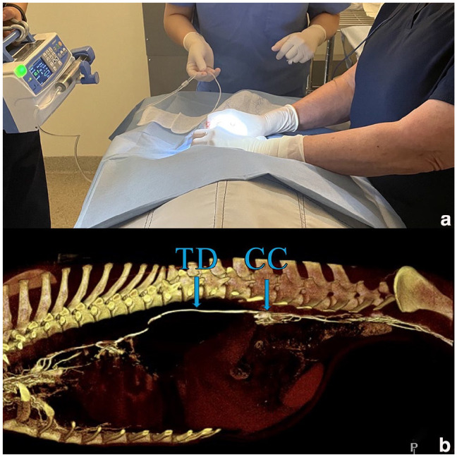
Case summary: A 4-year-old male castrated domestic shorthair cat presented with a 3-month history of coughing and progressive dyspnea. The cat was diagnosed with idiopathic chylothorax after serum biochemistry, thoracocentesis, cytology of pleural effusion, echocardiography and thoracic imaging were performed. After failure to respond to medical management with repeated thoracocenteses, the oral administration of rutin and a low-fat diet, the cat underwent staged interventions consisting of CT lymphangiogram, subtotal pericardiectomy and pleural access port (PAP) placement, followed by thoracic duct ligation (TDL) and cisterna chyli ablation (CCA) 2 months later. The cat made a clinical recovery based on resolution of chylous pleural effusion 1 month after TDL and CCA, and the PAP was removed 1 month later. The cat remained free of clinical signs 3 years postoperatively.
Relevance and novel information: This is the first report documenting the use of a PAP for the management of feline chylothorax to disease resolution. Employing this device enabled frequent, low morbidity drainage of the pleural effusion as an alternative to repeated thoracocenteses and facilitated staging the interventions throughout management of idiopathic chylothorax in a cat. PAPs can be used successfully in the management of chylous pleural effusion in cats.

 求助内容:
求助内容: 应助结果提醒方式:


