{"title":"Giant right atrial myxoma complicated with massive pulmonary embolism and right-sided heart failure: a case report.","authors":"Duolikun Mutailifu, Abudousaimi Aini, Abudunaibi Maimaitiaili","doi":"10.21037/acr-24-145","DOIUrl":null,"url":null,"abstract":"<p><strong>Background: </strong>About 75% of myxomas occur in the left atrium and 10-20% in the right atrium. Right atrium myxomas producing a wide range of potential symptoms and complications, from asymptomatic to serious presentations with acute pulmonary embolism (PE) or cardiogenic shock. Early diagnosis and prompt surgical intervention are critical.</p><p><strong>Case description: </strong>A 43-year-old man presented with progressive dyspnea, fatigue, and palpitations, imaging revealed a large mobile right atrial myxoma (78 mm × 52 mm) causing intermittent tricuspid valve obstruction and multifocal pulmonary emboli. Laboratory tests showed elevated B-type natriuretic peptide (BNP) and D-dimer levels. The patient was diagnosed with a benign cardiac tumor, PE, and New York Heart Association (NYHA) Class III heart function. Open-chest surgery with cardiopulmonary bypass resulted in successful tumor resection and emboli extraction. Postoperatively, the patient showed significant symptom improvement and no tumor recurrence at the 6-month follow-up. It is such giant right atrial myxomas that speak for the need for early diagnosis with surgical intervention.</p><p><strong>Conclusions: </strong>This case is illustrative of the practical outcome brought about by multidisciplinary involvement. These cases are rare, and the possibility of embolizing makes them inevitable for a meticulous clinical assessment with enhanced imaging, preferably with the help of computed tomography pulmonary angiography (CTPA) techniques, for diagnosis and management of these complex conditions. Early recognition and intervention are paramount to mitigating the risk of life-threatening complications such as pulmonary hypertension and embolism. Timely surgical intervention can prevent severe hemodynamic disturbances and improve patient outcomes.</p>","PeriodicalId":29752,"journal":{"name":"AME Case Reports","volume":"9 ","pages":"41"},"PeriodicalIF":0.7000,"publicationDate":"2025-01-06","publicationTypes":"Journal Article","fieldsOfStudy":null,"isOpenAccess":false,"openAccessPdf":"https://www.ncbi.nlm.nih.gov/pmc/articles/PMC12053975/pdf/","citationCount":"0","resultStr":null,"platform":"Semanticscholar","paperid":null,"PeriodicalName":"AME Case Reports","FirstCategoryId":"1085","ListUrlMain":"https://doi.org/10.21037/acr-24-145","RegionNum":0,"RegionCategory":null,"ArticlePicture":[],"TitleCN":null,"AbstractTextCN":null,"PMCID":null,"EPubDate":"2025/1/1 0:00:00","PubModel":"eCollection","JCR":"Q3","JCRName":"MEDICINE, GENERAL & INTERNAL","Score":null,"Total":0}
引用次数: 0
Abstract
Background: About 75% of myxomas occur in the left atrium and 10-20% in the right atrium. Right atrium myxomas producing a wide range of potential symptoms and complications, from asymptomatic to serious presentations with acute pulmonary embolism (PE) or cardiogenic shock. Early diagnosis and prompt surgical intervention are critical.
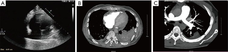
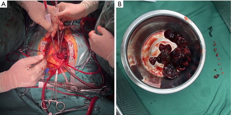
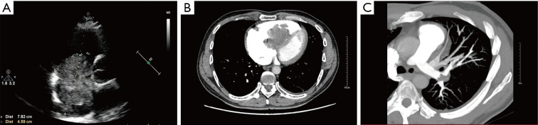
Case description: A 43-year-old man presented with progressive dyspnea, fatigue, and palpitations, imaging revealed a large mobile right atrial myxoma (78 mm × 52 mm) causing intermittent tricuspid valve obstruction and multifocal pulmonary emboli. Laboratory tests showed elevated B-type natriuretic peptide (BNP) and D-dimer levels. The patient was diagnosed with a benign cardiac tumor, PE, and New York Heart Association (NYHA) Class III heart function. Open-chest surgery with cardiopulmonary bypass resulted in successful tumor resection and emboli extraction. Postoperatively, the patient showed significant symptom improvement and no tumor recurrence at the 6-month follow-up. It is such giant right atrial myxomas that speak for the need for early diagnosis with surgical intervention.
Conclusions: This case is illustrative of the practical outcome brought about by multidisciplinary involvement. These cases are rare, and the possibility of embolizing makes them inevitable for a meticulous clinical assessment with enhanced imaging, preferably with the help of computed tomography pulmonary angiography (CTPA) techniques, for diagnosis and management of these complex conditions. Early recognition and intervention are paramount to mitigating the risk of life-threatening complications such as pulmonary hypertension and embolism. Timely surgical intervention can prevent severe hemodynamic disturbances and improve patient outcomes.

 求助内容:
求助内容: 应助结果提醒方式:


