{"title":"Navigating the Challenges of Persistent Left Superior Vena Cava in the Catheterization of Peripherally Inserted Central Catheter Port: A Case Study.","authors":"Takeshi Nakayama, Shinichiro Kobayashi, Shunsuke Murakami, Takahiro Enjoji, Hanako Tetsuo, Yusuke Inoue, Taichiro Kosaka, Akihiko Soyama, Tomohiko Adachi, Kazuma Kobayashi, Kengo Kanetaka, Susumu Eguchi","doi":"10.70352/scrj.cr.24-0088","DOIUrl":null,"url":null,"abstract":"<p><strong>Introduction: </strong>Persistent left superior vena cava (PLSVC), which is asymptomatic and occurs in 0.3%-0.5% of the general population, is typically detected incidentally but can complicate cardiac procedures owing to its potential to cause arrhythmias. This condition involves an additional venous return pathway to the right atrium, which can alter the cardiac anatomy and is associated with other cardiac aortic anomalies.</p><p><strong>Case presentation: </strong>A 75-year-old male patient required a central venous port for chemotherapy and radiation therapy for mid-thoracic esophageal cancer. Preoperative computed tomography images revealed that the PLSVC ran ventrally to the aortic and left pulmonary arteries, directly communicating with the right atrium. A peripherally inserted central catheter (PICC) port was planned. The catheter tip of the PICC port was placed within the left superior vena cava instead of the more common right superior vena cava, because the appropriate vessels could not be identified in the right upper arm. This anomaly necessitated a review of findings on the preoperative imaging and underscored the importance of early detection through echocardiography and radiographic guidance to prevent procedural complications. Reconstructed three-dimensional images and radiography-guided catheterization support the navigation of PICC port insertion.</p><p><strong>Conclusions: </strong>PLSVC, which is often asymptomatic, requires careful preprocedural planning and imaging to ensure safe PICC port insertion.</p>","PeriodicalId":22096,"journal":{"name":"Surgical Case Reports","volume":"11 1","pages":""},"PeriodicalIF":0.7000,"publicationDate":"2025-01-01","publicationTypes":"Journal Article","fieldsOfStudy":null,"isOpenAccess":false,"openAccessPdf":"https://www.ncbi.nlm.nih.gov/pmc/articles/PMC12055441/pdf/","citationCount":"0","resultStr":null,"platform":"Semanticscholar","paperid":null,"PeriodicalName":"Surgical Case Reports","FirstCategoryId":"1085","ListUrlMain":"https://doi.org/10.70352/scrj.cr.24-0088","RegionNum":0,"RegionCategory":null,"ArticlePicture":[],"TitleCN":null,"AbstractTextCN":null,"PMCID":null,"EPubDate":"2025/5/1 0:00:00","PubModel":"Epub","JCR":"Q4","JCRName":"SURGERY","Score":null,"Total":0}
引用次数: 0
Abstract
Introduction: Persistent left superior vena cava (PLSVC), which is asymptomatic and occurs in 0.3%-0.5% of the general population, is typically detected incidentally but can complicate cardiac procedures owing to its potential to cause arrhythmias. This condition involves an additional venous return pathway to the right atrium, which can alter the cardiac anatomy and is associated with other cardiac aortic anomalies.
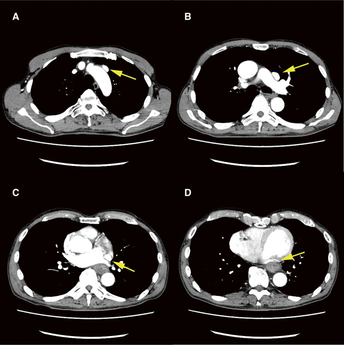
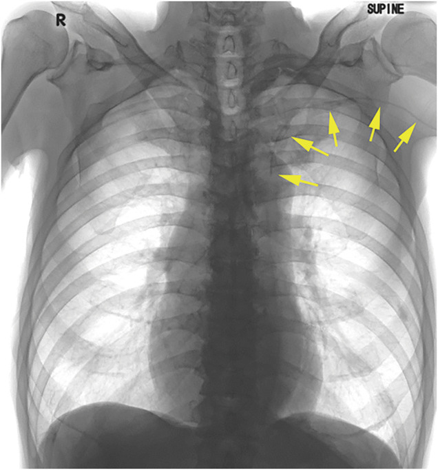
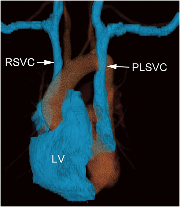
Case presentation: A 75-year-old male patient required a central venous port for chemotherapy and radiation therapy for mid-thoracic esophageal cancer. Preoperative computed tomography images revealed that the PLSVC ran ventrally to the aortic and left pulmonary arteries, directly communicating with the right atrium. A peripherally inserted central catheter (PICC) port was planned. The catheter tip of the PICC port was placed within the left superior vena cava instead of the more common right superior vena cava, because the appropriate vessels could not be identified in the right upper arm. This anomaly necessitated a review of findings on the preoperative imaging and underscored the importance of early detection through echocardiography and radiographic guidance to prevent procedural complications. Reconstructed three-dimensional images and radiography-guided catheterization support the navigation of PICC port insertion.
Conclusions: PLSVC, which is often asymptomatic, requires careful preprocedural planning and imaging to ensure safe PICC port insertion.

 求助内容:
求助内容: 应助结果提醒方式:


