{"title":"Evaluation of Mid-Palatal Suture Maturation Stage in Adolescents and Adults Using Cone Beam Computed Tomography (CBCT)- A Comparative Study.","authors":"Ravinder Singh, Deepak Gupta, Aashna Garg, Veenu Dahiya, Paras Gupta, Ramandeep Singh Gambhir","doi":"10.71480/nmj.v66i1.744","DOIUrl":null,"url":null,"abstract":"<p><strong>Background: </strong>Several methods have been described in orthodontics for the evaluation of the skeletal age. These include hand-wrist radiography and cervical vertebral maturation (CVM) based on lateral cephalogram. Computed tomography (CT) scan has emerged as an effective tool for image diagnosis in situ. The present study was done to assess the stages of the mid-palatal suture in adolescents and adults utilizing cone beam computed tomography (CBCT).</p><p><strong>Methodology: </strong>A descriptive and prospective study was done on 110 CBCT scans of individuals aged between 10-30 years, who visited the Department of Oral Medicine and Radiology, MMCDSR, Ambala, Haryana. The visualization and classification of the stage of maturation of the mid-palatine suture was done as per Angelieri's method using a cross-sectional axial slice.</p><p><strong>Results: </strong>Stage C was found to be the most prevalent (29.1%) with the majority of cases occurring in the 16-20 age range. Males were more likely to have Stage B, while females had Stage C. In 60 cases (54.5 percent of the overall sample), the mid-palatine suture was found to be open. The Chi-Square test results for each examiner were highly significant (p< 0.01), indicating a statistically significant association between age group and Stage distribution.</p><p><strong>Conclusion: </strong>There is a higher chance that post-adolescents and adults will have an open mid-palatal suture. When maxillary expansion is necessary, orthodontists may take these consequences into account. Furthermore, the middle palatal suture's ossification varies, hence using CBCT to rule out this possibility may be advised.</p>","PeriodicalId":94346,"journal":{"name":"Nigerian medical journal : journal of the Nigeria Medical Association","volume":"66 1","pages":"347-356"},"PeriodicalIF":0.0000,"publicationDate":"2025-04-03","publicationTypes":"Journal Article","fieldsOfStudy":null,"isOpenAccess":false,"openAccessPdf":"https://www.ncbi.nlm.nih.gov/pmc/articles/PMC12038629/pdf/","citationCount":"0","resultStr":null,"platform":"Semanticscholar","paperid":null,"PeriodicalName":"Nigerian medical journal : journal of the Nigeria Medical Association","FirstCategoryId":"1085","ListUrlMain":"https://doi.org/10.71480/nmj.v66i1.744","RegionNum":0,"RegionCategory":null,"ArticlePicture":[],"TitleCN":null,"AbstractTextCN":null,"PMCID":null,"EPubDate":"2025/1/1 0:00:00","PubModel":"eCollection","JCR":"","JCRName":"","Score":null,"Total":0}
引用次数: 0
Abstract
Background: Several methods have been described in orthodontics for the evaluation of the skeletal age. These include hand-wrist radiography and cervical vertebral maturation (CVM) based on lateral cephalogram. Computed tomography (CT) scan has emerged as an effective tool for image diagnosis in situ. The present study was done to assess the stages of the mid-palatal suture in adolescents and adults utilizing cone beam computed tomography (CBCT).
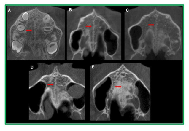
Methodology: A descriptive and prospective study was done on 110 CBCT scans of individuals aged between 10-30 years, who visited the Department of Oral Medicine and Radiology, MMCDSR, Ambala, Haryana. The visualization and classification of the stage of maturation of the mid-palatine suture was done as per Angelieri's method using a cross-sectional axial slice.
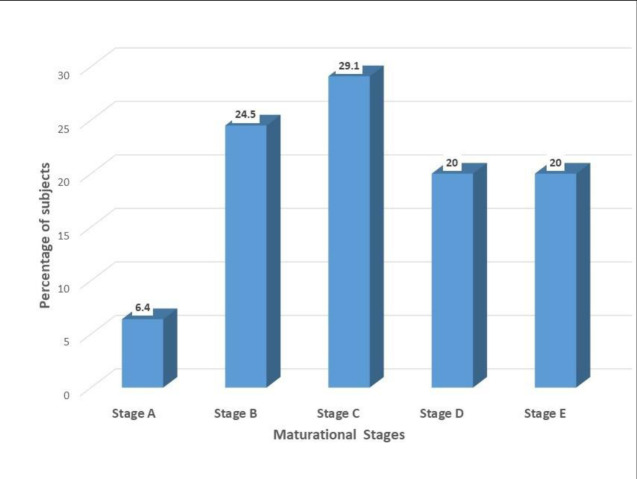
Results: Stage C was found to be the most prevalent (29.1%) with the majority of cases occurring in the 16-20 age range. Males were more likely to have Stage B, while females had Stage C. In 60 cases (54.5 percent of the overall sample), the mid-palatine suture was found to be open. The Chi-Square test results for each examiner were highly significant (p< 0.01), indicating a statistically significant association between age group and Stage distribution.
Conclusion: There is a higher chance that post-adolescents and adults will have an open mid-palatal suture. When maxillary expansion is necessary, orthodontists may take these consequences into account. Furthermore, the middle palatal suture's ossification varies, hence using CBCT to rule out this possibility may be advised.

 求助内容:
求助内容: 应助结果提醒方式:


