Utilization of 3D printing modeling techniques in the simulation instruction of ultrasound-guided puncture procedures on scoliotic spines of spinal muscular atrophy.
IF 3.1 Q1 RADIOLOGY, NUCLEAR MEDICINE & MEDICAL IMAGING
Di Xia, Fangliang Xing, Jiao Zhang, Jiaxin Lang, Gang Tan, Xulei Cui
{"title":"Utilization of 3D printing modeling techniques in the simulation instruction of ultrasound-guided puncture procedures on scoliotic spines of spinal muscular atrophy.","authors":"Di Xia, Fangliang Xing, Jiao Zhang, Jiaxin Lang, Gang Tan, Xulei Cui","doi":"10.1186/s41205-025-00266-x","DOIUrl":null,"url":null,"abstract":"<p><strong>Background: </strong>Puncture training with simulation models has emerged as a critical method for transmitting puncture skills, improving success rates, and minimizing injuries. Yet, obstacles such as proper material for ultrasound guidance, restricted options of 3D printing resources, and available substances to simulate human skin and muscle still hinder the production of simulation models that closely replicate clinical practice. This study aimed to develop a selective laser melting (SLM), 3D-printed simulation model that replicated the spine and skin contours of patients with spinal scoliosis.</p><p><strong>Methods: </strong>The 3D models of the scoliotic spines were developed from 3D reconstructions of high-resolution, computed tomography images from patients with spinal scoliosis, while the models of the skin to the bone structure were constructed based on the 3D reconstructions of the skin contours. SLM technology was used to print 3D models of the patients' spines. Gelatin casting was implemented to simulate the patients' skin and muscle tissues and to meet ultrasound anatomical requirements. Practical puncture training, which closely resembles clinical puncture practice, was then carried out to validate the effectiveness of the model. Improvements in proficiency and confidence in performing ultrasound-guided punctures after the simulation-model training were evaluated using the paired sample t test.</p><p><strong>Results: </strong>This research utilized 3D digital modeling, SLM 3D printing technology, and gelatin casting to establish simulation models of patients' spines and skin contours impacted by spinal scoliosis. The use of medical grade stainless steel material for modeling the spine and gelatin for skin and muscle tissues ensured the model had superior ultrasound anatomical properties. After the simulation training session, participants' proficiency and confidence in both ultrasound-assisted positioning and real-time guided puncture showed significant improvement, demonstrating the effectiveness of the simulation training model.</p><p><strong>Conclusions: </strong>The simulation model closely mimicked real clinical situations and was an effective training tool for medical professionals. Furthermore, these findings demonstrated the potential of 3D printing technology in developing simulation models that closely replicate real-world clinical scenarios and may have significant implications for medical education and training.</p>","PeriodicalId":72036,"journal":{"name":"3D printing in medicine","volume":"11 1","pages":"19"},"PeriodicalIF":3.1000,"publicationDate":"2025-04-27","publicationTypes":"Journal Article","fieldsOfStudy":null,"isOpenAccess":false,"openAccessPdf":"https://www.ncbi.nlm.nih.gov/pmc/articles/PMC12034200/pdf/","citationCount":"0","resultStr":null,"platform":"Semanticscholar","paperid":null,"PeriodicalName":"3D printing in medicine","FirstCategoryId":"1085","ListUrlMain":"https://doi.org/10.1186/s41205-025-00266-x","RegionNum":0,"RegionCategory":null,"ArticlePicture":[],"TitleCN":null,"AbstractTextCN":null,"PMCID":null,"EPubDate":"","PubModel":"","JCR":"Q1","JCRName":"RADIOLOGY, NUCLEAR MEDICINE & MEDICAL IMAGING","Score":null,"Total":0}
引用次数: 0
Abstract
Background: Puncture training with simulation models has emerged as a critical method for transmitting puncture skills, improving success rates, and minimizing injuries. Yet, obstacles such as proper material for ultrasound guidance, restricted options of 3D printing resources, and available substances to simulate human skin and muscle still hinder the production of simulation models that closely replicate clinical practice. This study aimed to develop a selective laser melting (SLM), 3D-printed simulation model that replicated the spine and skin contours of patients with spinal scoliosis.
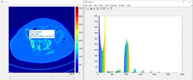
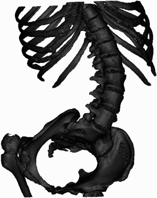
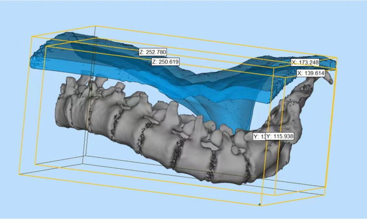
Methods: The 3D models of the scoliotic spines were developed from 3D reconstructions of high-resolution, computed tomography images from patients with spinal scoliosis, while the models of the skin to the bone structure were constructed based on the 3D reconstructions of the skin contours. SLM technology was used to print 3D models of the patients' spines. Gelatin casting was implemented to simulate the patients' skin and muscle tissues and to meet ultrasound anatomical requirements. Practical puncture training, which closely resembles clinical puncture practice, was then carried out to validate the effectiveness of the model. Improvements in proficiency and confidence in performing ultrasound-guided punctures after the simulation-model training were evaluated using the paired sample t test.
Results: This research utilized 3D digital modeling, SLM 3D printing technology, and gelatin casting to establish simulation models of patients' spines and skin contours impacted by spinal scoliosis. The use of medical grade stainless steel material for modeling the spine and gelatin for skin and muscle tissues ensured the model had superior ultrasound anatomical properties. After the simulation training session, participants' proficiency and confidence in both ultrasound-assisted positioning and real-time guided puncture showed significant improvement, demonstrating the effectiveness of the simulation training model.
Conclusions: The simulation model closely mimicked real clinical situations and was an effective training tool for medical professionals. Furthermore, these findings demonstrated the potential of 3D printing technology in developing simulation models that closely replicate real-world clinical scenarios and may have significant implications for medical education and training.

 求助内容:
求助内容: 应助结果提醒方式:


