Dionysios Adamopoulos, Georgios Rovas, Nicolas Johner, Hajo Müller, Jean-François Deux, Lindsey A. Crowe, Jean-Paul Vallée, François Mach, Nikolaos Stergiopulos, Dipen Shah
{"title":"Left atrial wall shear stress correlates with fibrosis in patients with atrial fibrillation","authors":"Dionysios Adamopoulos, Georgios Rovas, Nicolas Johner, Hajo Müller, Jean-François Deux, Lindsey A. Crowe, Jean-Paul Vallée, François Mach, Nikolaos Stergiopulos, Dipen Shah","doi":"10.1038/s44161-025-00651-z","DOIUrl":null,"url":null,"abstract":"Left atrial wall fibrosis has an important role in atrial fibrillation (AF) because of the abnormal electrophysiological properties of the fibrotic areas. However, the mechanisms behind the development of left atrial fibrosis are not well understood. Here, we examine the association between regional wall shear stress and areas with fibrosis in the left atrium of patients with AF. We recruited 15 patients with AF for an observational prospective study involving baseline three-dimensional (3D) electroanatomical mapping of the left atrium and preinterventional cardiovascular magnetic resonance imaging to detect left atrial fibrosis. We extracted a 3D anatomical model of the left atrium from the electroanatomical maps. Then, we calculated regional time-averaged wall shear stress (TAWSS) and blood stagnation by performing patient-specific computational fluid dynamic simulations. We found that fibrosis and electrical scarring were more prevalent in areas exposed to high TAWSS without blood stagnation, whereas areas with low TAWSS were associated with blood stagnation. Adamopoulos, Rovas et al. show that high regional wall shear stress correlates with fibrosis and electrical scarring in the left atrium of patients with atrial fibrillation, providing insight into the development of fibrotic tissue.","PeriodicalId":74245,"journal":{"name":"Nature cardiovascular research","volume":"4 6","pages":"677-688"},"PeriodicalIF":10.8000,"publicationDate":"2025-05-13","publicationTypes":"Journal Article","fieldsOfStudy":null,"isOpenAccess":false,"openAccessPdf":"https://www.ncbi.nlm.nih.gov/pmc/articles/PMC12170334/pdf/","citationCount":"0","resultStr":null,"platform":"Semanticscholar","paperid":null,"PeriodicalName":"Nature cardiovascular research","FirstCategoryId":"1085","ListUrlMain":"https://www.nature.com/articles/s44161-025-00651-z","RegionNum":0,"RegionCategory":null,"ArticlePicture":[],"TitleCN":null,"AbstractTextCN":null,"PMCID":null,"EPubDate":"","PubModel":"","JCR":"Q1","JCRName":"CARDIAC & CARDIOVASCULAR SYSTEMS","Score":null,"Total":0}
引用次数: 0
Abstract
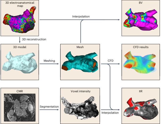
Left atrial wall fibrosis has an important role in atrial fibrillation (AF) because of the abnormal electrophysiological properties of the fibrotic areas. However, the mechanisms behind the development of left atrial fibrosis are not well understood. Here, we examine the association between regional wall shear stress and areas with fibrosis in the left atrium of patients with AF. We recruited 15 patients with AF for an observational prospective study involving baseline three-dimensional (3D) electroanatomical mapping of the left atrium and preinterventional cardiovascular magnetic resonance imaging to detect left atrial fibrosis. We extracted a 3D anatomical model of the left atrium from the electroanatomical maps. Then, we calculated regional time-averaged wall shear stress (TAWSS) and blood stagnation by performing patient-specific computational fluid dynamic simulations. We found that fibrosis and electrical scarring were more prevalent in areas exposed to high TAWSS without blood stagnation, whereas areas with low TAWSS were associated with blood stagnation. Adamopoulos, Rovas et al. show that high regional wall shear stress correlates with fibrosis and electrical scarring in the left atrium of patients with atrial fibrillation, providing insight into the development of fibrotic tissue.

 求助内容:
求助内容: 应助结果提醒方式:


