Prediction of Fluid Responsiveness Based on the External Jugular Vein Distensibility Index After Changes in Volume Status in Healthy, Anesthetized, and Mechanically Ventilated Dogs
Daeyun Seo, Seongsoo Lim, Beomkwan Namgoong, Heesung Uhm, Hyeajeong Hong, Nanju Lee, Isong Kim, Seunghun Heo, Ji Hwan Kang, Cheyoun Kim, Hayoung Shin, Jiwoong Her, Min-Su Kim
{"title":"Prediction of Fluid Responsiveness Based on the External Jugular Vein Distensibility Index After Changes in Volume Status in Healthy, Anesthetized, and Mechanically Ventilated Dogs","authors":"Daeyun Seo, Seongsoo Lim, Beomkwan Namgoong, Heesung Uhm, Hyeajeong Hong, Nanju Lee, Isong Kim, Seunghun Heo, Ji Hwan Kang, Cheyoun Kim, Hayoung Shin, Jiwoong Her, Min-Su Kim","doi":"10.1111/vec.13466","DOIUrl":null,"url":null,"abstract":"<div>\n \n \n <section>\n \n <h3> Objective</h3>\n \n <p>To investigate whether point-of-care ultrasound of the external jugular vein (EJV) can predict fluid responsiveness (FR) in healthy, anesthetized, mechanically ventilated dogs.</p>\n </section>\n \n <section>\n \n <h3> Design</h3>\n \n <p>Prospective, nonrandomized experimental study.</p>\n </section>\n \n <section>\n \n <h3> Setting</h3>\n \n <p>University-based small animal research facility.</p>\n </section>\n \n <section>\n \n <h3> Animals</h3>\n \n <p>Six healthy Beagle dogs.</p>\n </section>\n \n <section>\n \n <h3> Interventions</h3>\n \n <p>Dogs were investigated at six time points (TPs): baseline (TP<sub>1</sub>); 20 mL/kg of circulating blood was collected over 10 min (TP<sub>2</sub>); half of the collected blood was autotransfused for 10 min (TP<sub>3</sub>); remaining collected blood was autotransfused for 10 min (TP<sub>4</sub>); 0.9% normal saline (10 mL/kg for 10 min) was administered (TP<sub>5</sub>); and an additional dose of 0.9% normal saline (10 mL/kg for 10 min) was administered (TP<sub>6</sub>). Hemodynamic variables, Doppler images of the left ventricular outflow tract (LVOT), and M-mode images of the EJV were obtained at each TP. FR was evaluated during TP<sub>3–6</sub>. FR was defined as an increase of >15% in the LVOT velocity time integral following fluid challenge, while other results were defined as fluid nonresponsiveness (FNR). The external jugular vein distensibility index (EJVDI) was calculated as follows: [(maximal EJV diameter − minimal EJV diameter)/minimal EJV diameter] × 100%. The maximal EJV diameter was measured during inspiration, and the minimal EJV diameter was measured during expiration. In addition, gray zones indicating the range of diagnostic uncertainty were proposed in various indices for predicting FR.</p>\n </section>\n \n <section>\n \n <h3> Measurements and Main Results</h3>\n \n <p>Among the 24 fluid challenges performed between TP<sub>3</sub> and TP<sub>6</sub>, 11 FR and 13 FNR were identified. The area under the receiver operating characteristic curve for the EJVDI in predicting FR was 0.92, with a cut-ff value of 22.7%, and the gray zone was identified as 22.6%–27.3%.</p>\n </section>\n \n <section>\n \n <h3> Conclusions</h3>\n \n <p>The EJVDI could be used to predict FR in healthy, anesthetized, mechanically ventilated dogs. Further studies are required before point-of-care ultrasound of the EJV can be applied in various clinical settings.</p>\n </section>\n </div>","PeriodicalId":17603,"journal":{"name":"Journal of veterinary emergency and critical care","volume":"35 3","pages":"214-224"},"PeriodicalIF":1.2000,"publicationDate":"2025-04-29","publicationTypes":"Journal Article","fieldsOfStudy":null,"isOpenAccess":false,"openAccessPdf":"https://onlinelibrary.wiley.com/doi/epdf/10.1111/vec.13466","citationCount":"0","resultStr":null,"platform":"Semanticscholar","paperid":null,"PeriodicalName":"Journal of veterinary emergency and critical care","FirstCategoryId":"97","ListUrlMain":"https://onlinelibrary.wiley.com/doi/10.1111/vec.13466","RegionNum":3,"RegionCategory":"农林科学","ArticlePicture":[],"TitleCN":null,"AbstractTextCN":null,"PMCID":null,"EPubDate":"","PubModel":"","JCR":"Q3","JCRName":"VETERINARY SCIENCES","Score":null,"Total":0}
引用次数: 0
Abstract
Objective
To investigate whether point-of-care ultrasound of the external jugular vein (EJV) can predict fluid responsiveness (FR) in healthy, anesthetized, mechanically ventilated dogs.
Design
Prospective, nonrandomized experimental study.
Setting
University-based small animal research facility.
Animals
Six healthy Beagle dogs.
Interventions
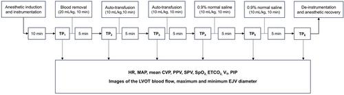
Dogs were investigated at six time points (TPs): baseline (TP1); 20 mL/kg of circulating blood was collected over 10 min (TP2); half of the collected blood was autotransfused for 10 min (TP3); remaining collected blood was autotransfused for 10 min (TP4); 0.9% normal saline (10 mL/kg for 10 min) was administered (TP5); and an additional dose of 0.9% normal saline (10 mL/kg for 10 min) was administered (TP6). Hemodynamic variables, Doppler images of the left ventricular outflow tract (LVOT), and M-mode images of the EJV were obtained at each TP. FR was evaluated during TP3–6. FR was defined as an increase of >15% in the LVOT velocity time integral following fluid challenge, while other results were defined as fluid nonresponsiveness (FNR). The external jugular vein distensibility index (EJVDI) was calculated as follows: [(maximal EJV diameter − minimal EJV diameter)/minimal EJV diameter] × 100%. The maximal EJV diameter was measured during inspiration, and the minimal EJV diameter was measured during expiration. In addition, gray zones indicating the range of diagnostic uncertainty were proposed in various indices for predicting FR.
Measurements and Main Results
Among the 24 fluid challenges performed between TP3 and TP6, 11 FR and 13 FNR were identified. The area under the receiver operating characteristic curve for the EJVDI in predicting FR was 0.92, with a cut-ff value of 22.7%, and the gray zone was identified as 22.6%–27.3%.
Conclusions
The EJVDI could be used to predict FR in healthy, anesthetized, mechanically ventilated dogs. Further studies are required before point-of-care ultrasound of the EJV can be applied in various clinical settings.
期刊介绍:
The Journal of Veterinary Emergency and Critical Care’s primary aim is to advance the international clinical standard of care for emergency/critical care patients of all species. The journal’s content is relevant to specialist and non-specialist veterinarians practicing emergency/critical care medicine. The journal achieves it aims by publishing descriptions of unique presentation or management; retrospective and prospective evaluations of prognosis, novel diagnosis, or therapy; translational basic science studies with clinical relevance; in depth reviews of pertinent topics; topical news and letters; and regular themed issues.
The journal is the official publication of the Veterinary Emergency and Critical Care Society, the American College of Veterinary Emergency and Critical Care, the European Veterinary Emergency and Critical Care Society, and the European College of Veterinary Emergency and Critical Care. It is a bimonthly publication with international impact and adheres to currently accepted ethical standards.
 求助内容:
求助内容: 应助结果提醒方式:


