{"title":"Downregulation of S100 calcium-binding A4 (S100A4) ameliorates hepatic fibrosis <i>via</i> regulating Wnt/β-catenin signaling pathway.","authors":"Chixian Zhang, Kai Bai, Dexu Li","doi":"10.4081/ejh.2025.4186","DOIUrl":null,"url":null,"abstract":"<p><p>S100 calcium-binding protein A4 (S100A4), a fibrosis-associated calcium-binding protein, has been implicated in fibrotic progression across multiple organs. Activation of the Wnt/β-catenin signaling pathway is a critical driver of hepatic fibrosis, yet the mechanistic role of S100A4 in this context remains poorly defined. This study investigated the regulatory role of S100A4 in hepatic fibrosis in vitro and in vivo. Hepatic stellate cells (HSCs) were treated with TGF-β to induce fibrotic activation, and S100A4 expression was silenced using shRNA. A carbon tetrachloride (CCl₄)-induced murine hepatic fibrosis model was employed for in vivo validation. Fibrotic markers, including collagen I, fibronectin, and α-smooth muscle actin (α-SMA), were assessed via qRT-PCR, Western blotting, immunofluorescence, and immunohistochemistry. Liver histopathology and function were evaluated using Masson trichrome staining, hematoxylin-eosin staining, and serum ALT/AST assays. In vitro experiments demonstrated that TGF-β treatment upregulated S100A4 expression in HSCs, while S100A4 silencing suppressed HSC activation, extracellular matrix (ECM) deposition, and Wnt/β-catenin signaling. In vivo, S100A4 downregulation attenuated CCl₄-induced hepatic fibrosis, reduced collagen accumulation, improved liver histology, and normalized serum ALT/AST levels. These findings indicate that S100A4 promotes hepatic fibrosis by activating the Wnt/β-catenin pathway, highlighting its potential as a therapeutic target.</p>","PeriodicalId":50487,"journal":{"name":"European Journal of Histochemistry","volume":"69 2","pages":""},"PeriodicalIF":2.1000,"publicationDate":"2025-04-07","publicationTypes":"Journal Article","fieldsOfStudy":null,"isOpenAccess":false,"openAccessPdf":"https://www.ncbi.nlm.nih.gov/pmc/articles/PMC12051413/pdf/","citationCount":"0","resultStr":null,"platform":"Semanticscholar","paperid":null,"PeriodicalName":"European Journal of Histochemistry","FirstCategoryId":"99","ListUrlMain":"https://doi.org/10.4081/ejh.2025.4186","RegionNum":4,"RegionCategory":"生物学","ArticlePicture":[],"TitleCN":null,"AbstractTextCN":null,"PMCID":null,"EPubDate":"2025/4/14 0:00:00","PubModel":"Epub","JCR":"Q4","JCRName":"CELL BIOLOGY","Score":null,"Total":0}
引用次数: 0
Abstract
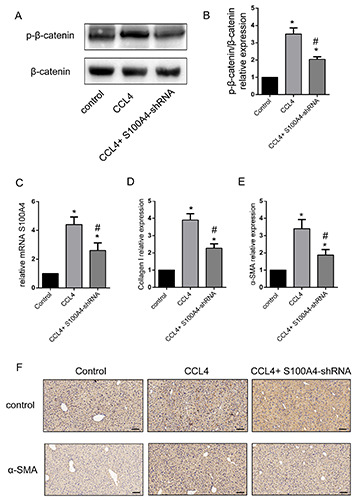
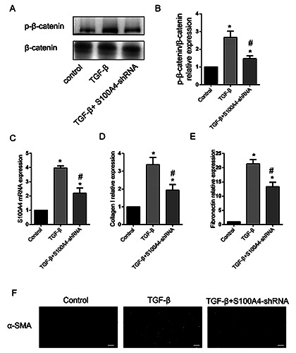
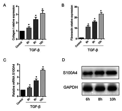
S100 calcium-binding protein A4 (S100A4), a fibrosis-associated calcium-binding protein, has been implicated in fibrotic progression across multiple organs. Activation of the Wnt/β-catenin signaling pathway is a critical driver of hepatic fibrosis, yet the mechanistic role of S100A4 in this context remains poorly defined. This study investigated the regulatory role of S100A4 in hepatic fibrosis in vitro and in vivo. Hepatic stellate cells (HSCs) were treated with TGF-β to induce fibrotic activation, and S100A4 expression was silenced using shRNA. A carbon tetrachloride (CCl₄)-induced murine hepatic fibrosis model was employed for in vivo validation. Fibrotic markers, including collagen I, fibronectin, and α-smooth muscle actin (α-SMA), were assessed via qRT-PCR, Western blotting, immunofluorescence, and immunohistochemistry. Liver histopathology and function were evaluated using Masson trichrome staining, hematoxylin-eosin staining, and serum ALT/AST assays. In vitro experiments demonstrated that TGF-β treatment upregulated S100A4 expression in HSCs, while S100A4 silencing suppressed HSC activation, extracellular matrix (ECM) deposition, and Wnt/β-catenin signaling. In vivo, S100A4 downregulation attenuated CCl₄-induced hepatic fibrosis, reduced collagen accumulation, improved liver histology, and normalized serum ALT/AST levels. These findings indicate that S100A4 promotes hepatic fibrosis by activating the Wnt/β-catenin pathway, highlighting its potential as a therapeutic target.
期刊介绍:
The Journal publishes original papers concerning investigations by histochemical and immunohistochemical methods, and performed with the aid of light, super-resolution and electron microscopy, cytometry and imaging techniques. Coverage extends to:
functional cell and tissue biology in animals and plants;
cell differentiation and death;
cell-cell interaction and molecular trafficking;
biology of cell development and senescence;
nerve and muscle cell biology;
cellular basis of diseases.
The histochemical approach is nowadays essentially aimed at locating molecules in the very place where they exert their biological roles, and at describing dynamically specific chemical activities in living cells. Basic research on cell functional organization is essential for understanding the mechanisms underlying major biological processes such as differentiation, the control of tissue homeostasis, and the regulation of normal and tumor cell growth. Even more than in the past, the European Journal of Histochemistry, as a journal of functional cytology, represents the venue where cell scientists may present and discuss their original results, technical improvements and theories.
 求助内容:
求助内容: 应助结果提醒方式:


