Ultrastructural and Immunohistochemical Study of a Gastric Bizarre Leiomyoma: Bizarre Nuclei and Prominent Cytoplasmic Processes Extending Into and Displacing the Stroma.
Kaoru Furihata, Waka Iwashita, Atsushi Kurabayashi, Kojima Koji, Mutsuo Furihata
{"title":"Ultrastructural and Immunohistochemical Study of a Gastric Bizarre Leiomyoma: Bizarre Nuclei and Prominent Cytoplasmic Processes Extending Into and Displacing the Stroma.","authors":"Kaoru Furihata, Waka Iwashita, Atsushi Kurabayashi, Kojima Koji, Mutsuo Furihata","doi":"10.1111/pin.70014","DOIUrl":null,"url":null,"abstract":"<p><p>This study presents the ultrastructural and immunohistochemical findings of a gastric bizarre leiomyoma arising in the vestibule of a 79-year-old male. Histologically, loosely proliferating tumor cells consist of large, multinucleated, bizarre nuclei with intranuclear inclusions and abundant cytoplasm-containing vacuoles. A murky line was apparent between the tumor cells and eosinophilic and heterogenous stroma-like areas. Immunohistochemically, tumor cells exhibited positively stained dot patterns of α-smooth muscle actin and caldesmon, which were distributed in the cytoplasm of tumor cells and stroma-like regions. Ultrastructurally, tumor cells exhibited extended and complex cytoplasmic processes comprising the fascicles of filamentous fibers. These structures were also detected in the apparent stroma-like regions observed histologically and were consistent with the α-smooth muscle actin- and caldesmon-immunopositive dot structures. The original stromal areas remained as considerably narrow gaps between tumor cells with extended cytoplasmic processes. To the best of our knowledge, this is the first report detailing the unique ultrastructural and immunohistochemical characteristics of tumor cells and the limited stromal composition of an extremely rare primary gastric bizarre leiomyoma.</p>","PeriodicalId":19806,"journal":{"name":"Pathology International","volume":" ","pages":"310-314"},"PeriodicalIF":3.4000,"publicationDate":"2025-06-01","publicationTypes":"Journal Article","fieldsOfStudy":null,"isOpenAccess":false,"openAccessPdf":"https://www.ncbi.nlm.nih.gov/pmc/articles/PMC12184308/pdf/","citationCount":"0","resultStr":null,"platform":"Semanticscholar","paperid":null,"PeriodicalName":"Pathology International","FirstCategoryId":"3","ListUrlMain":"https://doi.org/10.1111/pin.70014","RegionNum":4,"RegionCategory":"医学","ArticlePicture":[],"TitleCN":null,"AbstractTextCN":null,"PMCID":null,"EPubDate":"2025/4/14 0:00:00","PubModel":"Epub","JCR":"Q2","JCRName":"PATHOLOGY","Score":null,"Total":0}
引用次数: 0
Abstract
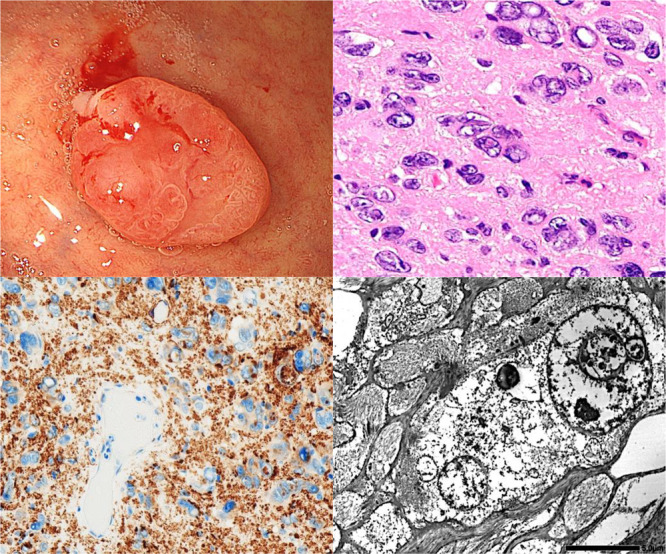
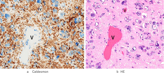
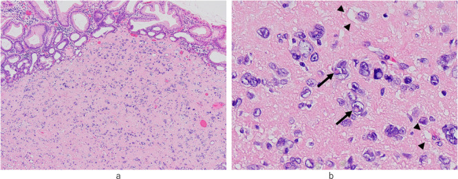
This study presents the ultrastructural and immunohistochemical findings of a gastric bizarre leiomyoma arising in the vestibule of a 79-year-old male. Histologically, loosely proliferating tumor cells consist of large, multinucleated, bizarre nuclei with intranuclear inclusions and abundant cytoplasm-containing vacuoles. A murky line was apparent between the tumor cells and eosinophilic and heterogenous stroma-like areas. Immunohistochemically, tumor cells exhibited positively stained dot patterns of α-smooth muscle actin and caldesmon, which were distributed in the cytoplasm of tumor cells and stroma-like regions. Ultrastructurally, tumor cells exhibited extended and complex cytoplasmic processes comprising the fascicles of filamentous fibers. These structures were also detected in the apparent stroma-like regions observed histologically and were consistent with the α-smooth muscle actin- and caldesmon-immunopositive dot structures. The original stromal areas remained as considerably narrow gaps between tumor cells with extended cytoplasmic processes. To the best of our knowledge, this is the first report detailing the unique ultrastructural and immunohistochemical characteristics of tumor cells and the limited stromal composition of an extremely rare primary gastric bizarre leiomyoma.
期刊介绍:
Pathology International is the official English journal of the Japanese Society of Pathology, publishing articles of excellence in human and experimental pathology. The Journal focuses on the morphological study of the disease process and/or mechanisms. For human pathology, morphological investigation receives priority but manuscripts describing the result of any ancillary methods (cellular, chemical, immunological and molecular biological) that complement the morphology are accepted. Manuscript on experimental pathology that approach pathologenesis or mechanisms of disease processes are expected to report on the data obtained from models using cellular, biochemical, molecular biological, animal, immunological or other methods in conjunction with morphology. Manuscripts that report data on laboratory medicine (clinical pathology) without significant morphological contribution are not accepted.

 求助内容:
求助内容: 应助结果提醒方式:


