{"title":"Unveiling HER2 immunoexpression in canine hepatoid gland neoplasms: clinicopathological and morphological associations.","authors":"Hassadin Boonsriroj, Sahatchai Tangtrongsup, Jirapat Arunorat, Thanongsak Mamom, Pinkarn Chantawong","doi":"10.1080/23144599.2025.2495522","DOIUrl":null,"url":null,"abstract":"<p><p>Canine hepatoid gland neoplasms (HGNs) are significant clinical concerns due to their high prevalence and diverse biological behaviour. Human epidermal growth factor receptor 2 (HER2), a tyrosine kinase receptor implicated in various aspects of tumorigenesis, has been extensively studied in human and animal neoplasms but remains unexplored in HGNs. This study aimed to assess HER2 immunoexpression in canine HGNs and its association with clinicopathological and morphological features. A total of 61 formalin-fixed paraffin-embedded samples, including normal hepatoid glands (<i>n</i> = 10), hepatoid gland adenomas (HGAs, <i>n</i> = 20), hepatoid gland epitheliomas (HGEs, <i>n</i> = 16), and hepatoid gland carcinomas (HGCs, <i>n</i> = 15), were analysed using immunohistochemistry. HER2 expression was scored based on percentage positivity and staining intensity. HER2-positive expression was detected in 50% of HGEs (score 2 + ) and 73.3% of HGCs, with 36.4% of cases scoring 3 + . In contrast, all HGAs and normal hepatoid tissues were HER2-immunonegative. Statistical analysis revealed significant differences in HER2 expression among normal and neoplastic hepatoid glands (<i>p</i> < 0.001). Only in HGCs, HER2 expression was significantly associated with tissue invasion (<i>p</i> = 0.007), mitotic count (<i>p</i> = 0.033), and nuclear pleomorphism (<i>p</i> = 0.007). These findings suggest that HER2 may play a role in the progression of malignant HGNs, particularly HGCs. This preliminary study highlights the potential of HER2 as a diagnostic marker and emphasizes the need for further investigation into its prognostic value and role in HER2-targeted therapy for canine HGCs.</p>","PeriodicalId":45744,"journal":{"name":"International Journal of Veterinary Science and Medicine","volume":"13 1","pages":"1-12"},"PeriodicalIF":3.2000,"publicationDate":"2025-04-24","publicationTypes":"Journal Article","fieldsOfStudy":null,"isOpenAccess":false,"openAccessPdf":"https://www.ncbi.nlm.nih.gov/pmc/articles/PMC12024500/pdf/","citationCount":"0","resultStr":null,"platform":"Semanticscholar","paperid":null,"PeriodicalName":"International Journal of Veterinary Science and Medicine","FirstCategoryId":"1085","ListUrlMain":"https://doi.org/10.1080/23144599.2025.2495522","RegionNum":0,"RegionCategory":null,"ArticlePicture":[],"TitleCN":null,"AbstractTextCN":null,"PMCID":null,"EPubDate":"2025/1/1 0:00:00","PubModel":"eCollection","JCR":"Q1","JCRName":"VETERINARY SCIENCES","Score":null,"Total":0}
引用次数: 0
Abstract
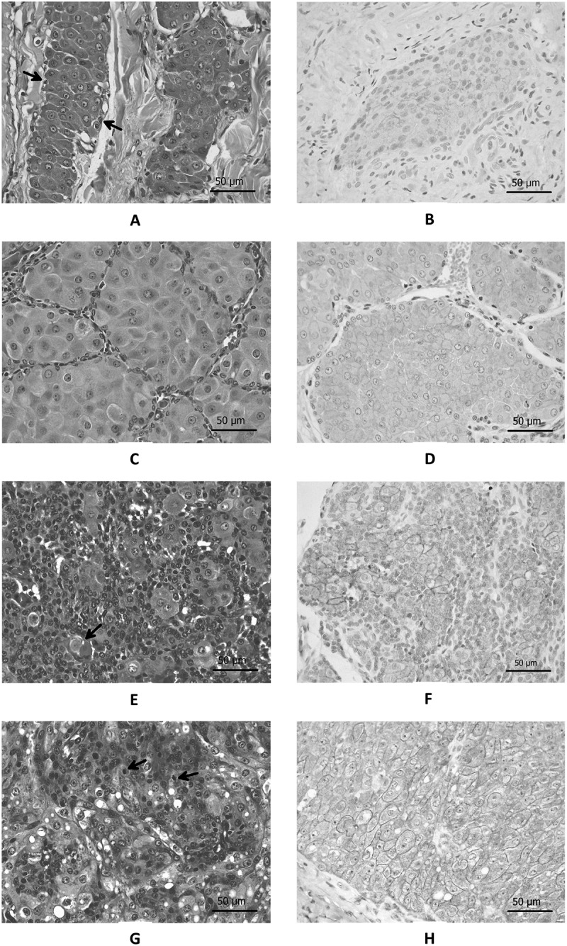
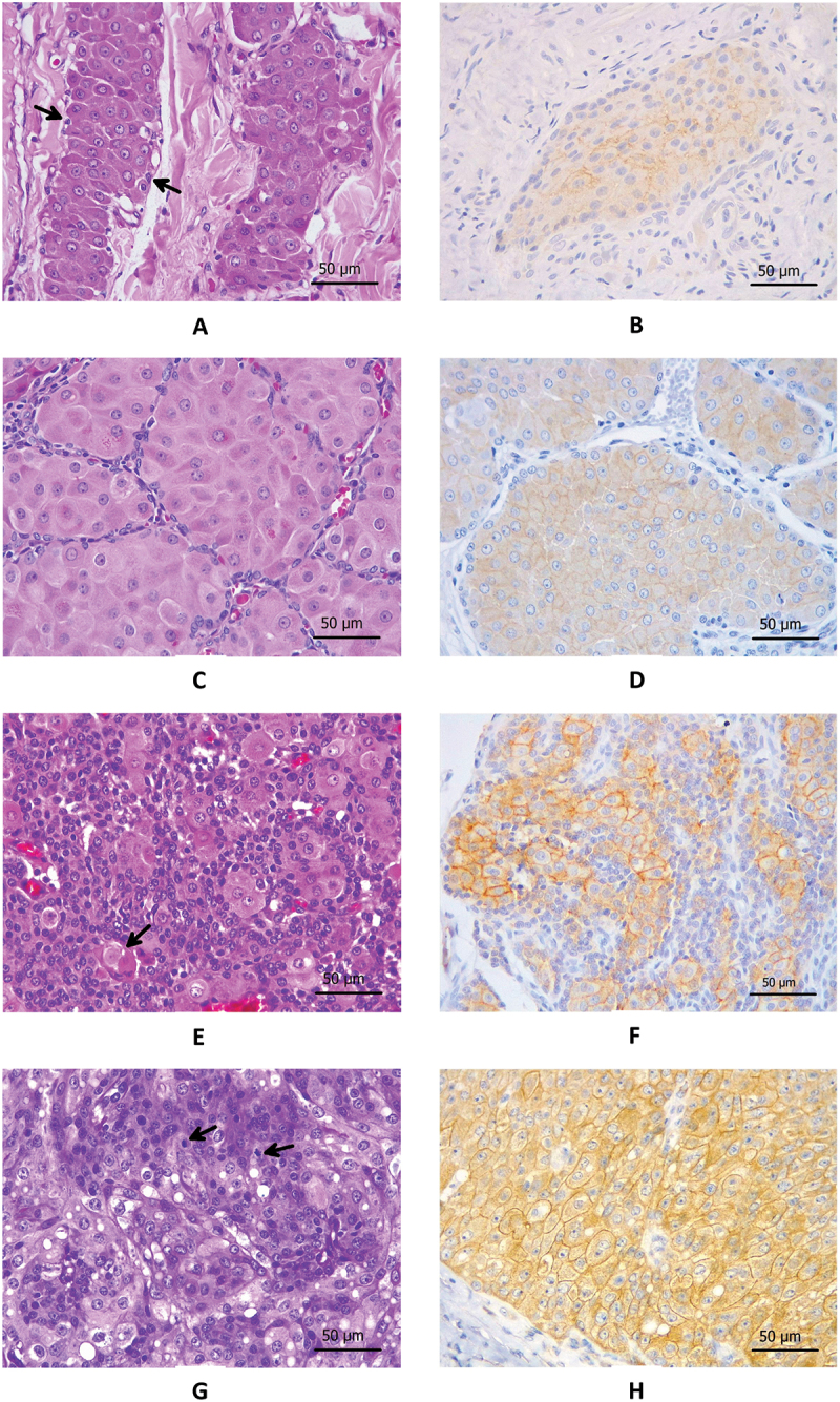
Canine hepatoid gland neoplasms (HGNs) are significant clinical concerns due to their high prevalence and diverse biological behaviour. Human epidermal growth factor receptor 2 (HER2), a tyrosine kinase receptor implicated in various aspects of tumorigenesis, has been extensively studied in human and animal neoplasms but remains unexplored in HGNs. This study aimed to assess HER2 immunoexpression in canine HGNs and its association with clinicopathological and morphological features. A total of 61 formalin-fixed paraffin-embedded samples, including normal hepatoid glands (n = 10), hepatoid gland adenomas (HGAs, n = 20), hepatoid gland epitheliomas (HGEs, n = 16), and hepatoid gland carcinomas (HGCs, n = 15), were analysed using immunohistochemistry. HER2 expression was scored based on percentage positivity and staining intensity. HER2-positive expression was detected in 50% of HGEs (score 2 + ) and 73.3% of HGCs, with 36.4% of cases scoring 3 + . In contrast, all HGAs and normal hepatoid tissues were HER2-immunonegative. Statistical analysis revealed significant differences in HER2 expression among normal and neoplastic hepatoid glands (p < 0.001). Only in HGCs, HER2 expression was significantly associated with tissue invasion (p = 0.007), mitotic count (p = 0.033), and nuclear pleomorphism (p = 0.007). These findings suggest that HER2 may play a role in the progression of malignant HGNs, particularly HGCs. This preliminary study highlights the potential of HER2 as a diagnostic marker and emphasizes the need for further investigation into its prognostic value and role in HER2-targeted therapy for canine HGCs.

 求助内容:
求助内容: 应助结果提醒方式:


