Andrada-Elena Mirescu, Dan George Deleanu, George Baltă, Ioana Teodora Tofolean, Florian Baltă, Irina-Elena Cristescu, Sanda Jurja
{"title":"Retinal Vascular Diseases Highlighted by Adaptive Optics Ophthalmoscopy.","authors":"Andrada-Elena Mirescu, Dan George Deleanu, George Baltă, Ioana Teodora Tofolean, Florian Baltă, Irina-Elena Cristescu, Sanda Jurja","doi":"10.22336/rjo.2025.07","DOIUrl":null,"url":null,"abstract":"<p><strong>Objective: </strong>We used adaptive optics ophthalmoscopy to assess retinal microcirculation and photoreceptor parameters in healthy individuals and patients with vascular retinal diseases. This technology enhances optical system resolution to 2 µm by correcting wavefront aberrations, revolutionizing in vivo studies of ocular structures.</p><p><strong>Methods: </strong>Our study examined the clinical applications of adaptive optics in both healthy individuals and patients with vascular retinal diseases, including nonproliferative diabetic retinopathy, proliferative diabetic retinopathy, and macular telangiectasia (MacTel) type 2.</p><p><strong>Results: </strong>Our study underlined a higher wall-to-lumen ratio (WLR) value in our patient with proliferative diabetic retinopathy compared to our healthy volunteer. Additionally, we found a positive correlation between WLR and the severity of diabetic retinopathy. Furthermore, cone density was lower in all quadrants with proliferative diabetic retinopathy. For our patient diagnosed with MacTel type 2, the cone mosaic appeared irregular and blurred, with notable cone loss, especially on the temporal side of the macula, consistent with the typical location of MacTel type 2 lesions.</p><p><strong>Discussion: </strong>Adaptive optics imaging assesses retinal changes in vascular diseases despite acquisition challenges. The obtained images aid in tracking diabetic retinopathy progression and detecting early MacTel Type 2 changes. Our study highlighted vascular and photoreceptor changes, quantifying these parameters to enhance understanding of these vascular diseases.</p><p><strong>Conclusions: </strong>Adaptive optics imaging is an advanced technique that provides high-resolution visualization of the microstructure of retinal vasculature and photoreceptors. This technology enhances our understanding of healthy and vascular retinal conditions, aiding diagnosis, monitoring, and prognosis.</p>","PeriodicalId":94355,"journal":{"name":"Romanian journal of ophthalmology","volume":"69 1","pages":"36-41"},"PeriodicalIF":0.0000,"publicationDate":"2025-01-01","publicationTypes":"Journal Article","fieldsOfStudy":null,"isOpenAccess":false,"openAccessPdf":"https://www.ncbi.nlm.nih.gov/pmc/articles/PMC12049653/pdf/","citationCount":"0","resultStr":null,"platform":"Semanticscholar","paperid":null,"PeriodicalName":"Romanian journal of ophthalmology","FirstCategoryId":"1085","ListUrlMain":"https://doi.org/10.22336/rjo.2025.07","RegionNum":0,"RegionCategory":null,"ArticlePicture":[],"TitleCN":null,"AbstractTextCN":null,"PMCID":null,"EPubDate":"","PubModel":"","JCR":"","JCRName":"","Score":null,"Total":0}
引用次数: 0
Abstract
Objective: We used adaptive optics ophthalmoscopy to assess retinal microcirculation and photoreceptor parameters in healthy individuals and patients with vascular retinal diseases. This technology enhances optical system resolution to 2 µm by correcting wavefront aberrations, revolutionizing in vivo studies of ocular structures.
Methods: Our study examined the clinical applications of adaptive optics in both healthy individuals and patients with vascular retinal diseases, including nonproliferative diabetic retinopathy, proliferative diabetic retinopathy, and macular telangiectasia (MacTel) type 2.
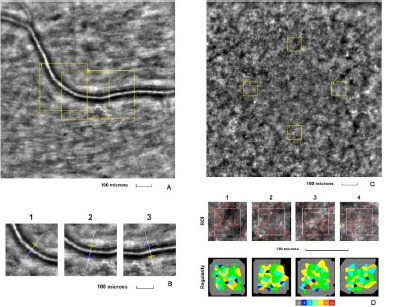
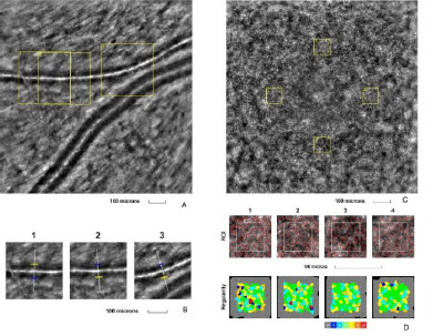
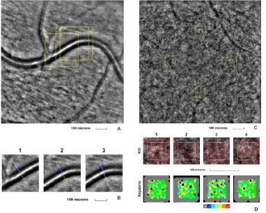
Results: Our study underlined a higher wall-to-lumen ratio (WLR) value in our patient with proliferative diabetic retinopathy compared to our healthy volunteer. Additionally, we found a positive correlation between WLR and the severity of diabetic retinopathy. Furthermore, cone density was lower in all quadrants with proliferative diabetic retinopathy. For our patient diagnosed with MacTel type 2, the cone mosaic appeared irregular and blurred, with notable cone loss, especially on the temporal side of the macula, consistent with the typical location of MacTel type 2 lesions.
Discussion: Adaptive optics imaging assesses retinal changes in vascular diseases despite acquisition challenges. The obtained images aid in tracking diabetic retinopathy progression and detecting early MacTel Type 2 changes. Our study highlighted vascular and photoreceptor changes, quantifying these parameters to enhance understanding of these vascular diseases.
Conclusions: Adaptive optics imaging is an advanced technique that provides high-resolution visualization of the microstructure of retinal vasculature and photoreceptors. This technology enhances our understanding of healthy and vascular retinal conditions, aiding diagnosis, monitoring, and prognosis.

 求助内容:
求助内容: 应助结果提醒方式:


