Baris Esen, Okan Falay, Kayhan Tarim, Hulya Seymen, Mert Kilic, Sevil Bavbek, Yakup Kordan, Mehmet Onur Demirkol, Derya Tilki, Tarik Esen
{"title":"Revisiting Skull Metastases of Prostate Cancer at Prostate-Specific Membrane Antigen (PSMA) Positron Emission Tomography/Computed Tomography Era: PSMA Uptake Characteristics and Oncological Outcomes.","authors":"Baris Esen, Okan Falay, Kayhan Tarim, Hulya Seymen, Mert Kilic, Sevil Bavbek, Yakup Kordan, Mehmet Onur Demirkol, Derya Tilki, Tarik Esen","doi":"10.5152/tud.2025.24164","DOIUrl":null,"url":null,"abstract":"<p><strong>Objective: </strong>We aimed to evaluate prostate-specific membrane antigen (PSMA) uptake characteristics and the oncological outcomes in patients with skull metastases.</p><p><strong>Methods: </strong>The records of 345 serial PSMA positron emission tomography (PET)/computed tomography (CT) scans of 96 patients with metastatic prostate cancer (PCa) were evaluated retrospectively. Skull bone metastasis was detected in 18 patients (18/96, 18.7%), with a mean age of 72.4 ± 9.1 years, and in 40 PSMA PET/CT scans (40/345, 11.6%). Involved skull bones, PSMA uptake characteristics, and CT counterparts of metastatic lesions were centrally reviewed. Prostate specific antigen (PSA) levels at the time of skull metastasis detection and PSMA-detected other metastatic lesions were recorded.</p><p><strong>Results: </strong>All patients with a skull metastasis showed multiple other metastatic bone lesions, and 6 (33.3%) had visceral metastasis. Seven (38.9%) patients had solitary skull lesions, whereas 11 (61.1%) had multiple skull metastases. Twenty-two out of 37 (59.5%) metastatic lesions had no CT counterpart. The median SUVmax was significantly higher in metastatic lesions with a CT counterpart (median 9.09 vs. 4.63, P = .018). At a median follow-up of 23.4 mo (interquartile range [IQR] 8.7-34.1) after detection of skull metastasis, 5 out of 11 (45.5%) hormone-sensitive and all castration-resistant patients died of PCa. The median survival of patients with castration-resistant disease was 9.92 months.</p><p><strong>Conclusion: </strong>The majority of PSMA-detected skull metastases did not show a CT counterpart, which may explain why skull metastases were rarely detected before the PSMA PET-era. In high-volume metastatic prostatic cancer cases, 68Ga-PSMA PET/CT imaging field including the vertex, may enhance the accuracy in detecting tumor extent and metabolic tumor volume.</p>","PeriodicalId":101337,"journal":{"name":"Urology research & practice","volume":"50 5","pages":"275-280"},"PeriodicalIF":1.1000,"publicationDate":"2025-03-07","publicationTypes":"Journal Article","fieldsOfStudy":null,"isOpenAccess":false,"openAccessPdf":"https://www.ncbi.nlm.nih.gov/pmc/articles/PMC11923601/pdf/","citationCount":"0","resultStr":null,"platform":"Semanticscholar","paperid":null,"PeriodicalName":"Urology research & practice","FirstCategoryId":"1085","ListUrlMain":"https://doi.org/10.5152/tud.2025.24164","RegionNum":0,"RegionCategory":null,"ArticlePicture":[],"TitleCN":null,"AbstractTextCN":null,"PMCID":null,"EPubDate":"","PubModel":"","JCR":"0","JCRName":"UROLOGY & NEPHROLOGY","Score":null,"Total":0}
引用次数: 0
Abstract
Objective: We aimed to evaluate prostate-specific membrane antigen (PSMA) uptake characteristics and the oncological outcomes in patients with skull metastases.
Methods: The records of 345 serial PSMA positron emission tomography (PET)/computed tomography (CT) scans of 96 patients with metastatic prostate cancer (PCa) were evaluated retrospectively. Skull bone metastasis was detected in 18 patients (18/96, 18.7%), with a mean age of 72.4 ± 9.1 years, and in 40 PSMA PET/CT scans (40/345, 11.6%). Involved skull bones, PSMA uptake characteristics, and CT counterparts of metastatic lesions were centrally reviewed. Prostate specific antigen (PSA) levels at the time of skull metastasis detection and PSMA-detected other metastatic lesions were recorded.
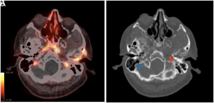
Results: All patients with a skull metastasis showed multiple other metastatic bone lesions, and 6 (33.3%) had visceral metastasis. Seven (38.9%) patients had solitary skull lesions, whereas 11 (61.1%) had multiple skull metastases. Twenty-two out of 37 (59.5%) metastatic lesions had no CT counterpart. The median SUVmax was significantly higher in metastatic lesions with a CT counterpart (median 9.09 vs. 4.63, P = .018). At a median follow-up of 23.4 mo (interquartile range [IQR] 8.7-34.1) after detection of skull metastasis, 5 out of 11 (45.5%) hormone-sensitive and all castration-resistant patients died of PCa. The median survival of patients with castration-resistant disease was 9.92 months.
Conclusion: The majority of PSMA-detected skull metastases did not show a CT counterpart, which may explain why skull metastases were rarely detected before the PSMA PET-era. In high-volume metastatic prostatic cancer cases, 68Ga-PSMA PET/CT imaging field including the vertex, may enhance the accuracy in detecting tumor extent and metabolic tumor volume.


 求助内容:
求助内容: 应助结果提醒方式:


