{"title":"Histopathological and molecular analysis in dermis and epidermis of patients with systemic and localized scleroderma.","authors":"Betul Sozeri, Seyma Turkmen, Basak Yildiz Atikan, Huseyin Aktug","doi":"10.14744/nci.2024.45389","DOIUrl":null,"url":null,"abstract":"<p><strong>Objective: </strong>Scleroderma has a wide range of clinical manifestations due to vasculopathy, autoimmunity, altered endothelium function, and abnormal fibrosis, which are accused in the pathogenesis of the disease. The aim of this study is to shed light on the pathogenesis of the disease in childhood via dermal immunohistochemical analysis of the cases.</p><p><strong>Methods: </strong>A single-blind clinical trial is conducted with evaluation of the tissue samples obtained from patients. The samples are stained with PAS, hematoxylin and eosin, E-Cadherin, Connective tissue growth factor (CTGF), Tunnel, and staining for Transforming growth factor beta 1 (TGF-β1) and evaluated by light microscopy. In addition, both TGF-β1 level and mRNA expression analyses in plasma and tissue samples from patients are performed. A total of 15 patients (systemic, n=8 or localized; n=7) were enrolled in the study.</p><p><strong>Results: </strong>The mean age of onset of the disease was 9.2±1.2 years, and the mean age of diagnosis was 15.3±3.2 years. Antinuclear antibody (ANA) titer was between 1/160-1/640 in all patients with systemic sclerosis. There was no ANA positivity in patients with localized scleroderma. A total of 22 tissue samples (15 diseased tissues, 7 healthy tissues) were examined. Histopathological examination has shown that two clinically different subgroups have different characteristics at the tissue level.</p><p><strong>Conclusion: </strong>TGF-β1 levels, which play a fundamental role in the pathogenesis of the disease, are found in both plasma and skin have been shown high. This elevation was found particularly in patients with systemic scleroderma to be more pronounced. Also, in patients with localized scleroderma, skin fibroblasts have been shown to limit the pathologic response.</p>","PeriodicalId":94347,"journal":{"name":"Northern clinics of Istanbul","volume":"12 2","pages":"189-195"},"PeriodicalIF":0.9000,"publicationDate":"2025-04-22","publicationTypes":"Journal Article","fieldsOfStudy":null,"isOpenAccess":false,"openAccessPdf":"https://www.ncbi.nlm.nih.gov/pmc/articles/PMC12050995/pdf/","citationCount":"0","resultStr":null,"platform":"Semanticscholar","paperid":null,"PeriodicalName":"Northern clinics of Istanbul","FirstCategoryId":"1085","ListUrlMain":"https://doi.org/10.14744/nci.2024.45389","RegionNum":0,"RegionCategory":null,"ArticlePicture":[],"TitleCN":null,"AbstractTextCN":null,"PMCID":null,"EPubDate":"2025/1/1 0:00:00","PubModel":"eCollection","JCR":"","JCRName":"","Score":null,"Total":0}
引用次数: 0
Abstract
Objective: Scleroderma has a wide range of clinical manifestations due to vasculopathy, autoimmunity, altered endothelium function, and abnormal fibrosis, which are accused in the pathogenesis of the disease. The aim of this study is to shed light on the pathogenesis of the disease in childhood via dermal immunohistochemical analysis of the cases.
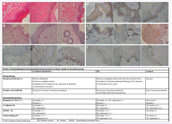
Methods: A single-blind clinical trial is conducted with evaluation of the tissue samples obtained from patients. The samples are stained with PAS, hematoxylin and eosin, E-Cadherin, Connective tissue growth factor (CTGF), Tunnel, and staining for Transforming growth factor beta 1 (TGF-β1) and evaluated by light microscopy. In addition, both TGF-β1 level and mRNA expression analyses in plasma and tissue samples from patients are performed. A total of 15 patients (systemic, n=8 or localized; n=7) were enrolled in the study.
Results: The mean age of onset of the disease was 9.2±1.2 years, and the mean age of diagnosis was 15.3±3.2 years. Antinuclear antibody (ANA) titer was between 1/160-1/640 in all patients with systemic sclerosis. There was no ANA positivity in patients with localized scleroderma. A total of 22 tissue samples (15 diseased tissues, 7 healthy tissues) were examined. Histopathological examination has shown that two clinically different subgroups have different characteristics at the tissue level.
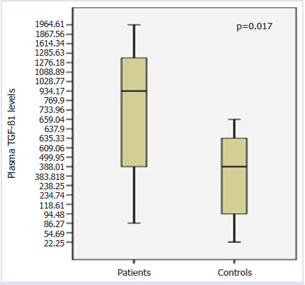
Conclusion: TGF-β1 levels, which play a fundamental role in the pathogenesis of the disease, are found in both plasma and skin have been shown high. This elevation was found particularly in patients with systemic scleroderma to be more pronounced. Also, in patients with localized scleroderma, skin fibroblasts have been shown to limit the pathologic response.

 求助内容:
求助内容: 应助结果提醒方式:


