{"title":"Analysis of mast cells in dental follicle and dentigerous cyst: A histopathological study.","authors":"Jayaraman Sindhumati, Sanjai Karpagaselvi, Kumaraswamy Jayalakshmi, Papaiah Lokesh, Keshavaiah Roopavathi, Pandey Bhavna","doi":"10.4103/jomfp.jomfp_479_23","DOIUrl":null,"url":null,"abstract":"<p><strong>Introduction: </strong>Mast cells are large granular cells that arise from multipotent CD 34+ precursors in the bone marrow normally distributed throughout the connective tissues. Following activation of immunologic or nonimmunologic stimuli, mast cells release secretory granules which give the characteristic metachromatic appearance with toluidine blue stain. Release of numerous mediators on degranulation of mast cells plays an important role in the pathogenesis of odontogenic cysts.</p><p><strong>Context: </strong>Odontogenic cysts, such as dentigerous cysts, arise due to the accumulation of fluid between the crown of an unerupted tooth and the reduced enamel epithelium. Dental follicles, which surround developing teeth, can also undergo cystic transformation. Mast cells activity might contribute to cyst expansion and bone resorption, highlighting their potential role in cystic pathology.</p><p><strong>Aims: </strong>To study the presence of mast cells in the dental follicle and dentigerous cyst. To quantify the mast cells in the abovementioned subjects. To study the pattern of distribution of mast cell distribution in different zones of the study groups.</p><p><strong>Settings and design: </strong>This was histopathological study conducted at the Department of Oral Pathology and Microbiology, Vydehi College of Dental Sciences and Research Centre, Bengaluru, between 2012 and 2015.</p><p><strong>Methods and material: </strong>Our study was conducted in the Department of Oral Pathology and Microbiology at Vydehi College of Dental Sciences and Research Centre in the year 2012 to 2015. Histopathologically analyzed 30 cases each of dental follicle, and dentigerous cysts were taken and 4-5 micron sections were stained with toluidine blue. Counting of mast cells was done in three different zones which included subepithelial, intermediate, and deep zone. The results were tabulated and statistically analyzed.</p><p><strong>Statistical analysis used: </strong>Kruskal-Wallis Chi-squared test.</p><p><strong>Results: </strong>Both dental follicles and dentigerous cysts showed the presence of mast cells, and highest numbers of mast cells were seen in subepithelial zone followed by intermediate and deep zones. There was statistically significant relation in the number of mast cells in dentigerous cysts and dental follicle along subepithelial and intermediate zone with a <i>P</i> value of <0.05. In our study, we also found increased mast cell count in inflamed cases of dental follicle and dentigerous cyst compared with noninflamed cases with a <i>P</i> value of <0.01.</p><p><strong>Conclusions: </strong>It is well known that mast cells play a role in the initiation of inflammation, and this inflammatory process may be associated with pericoronal follicle enlargement, a process that could result in cystic transformation of the follicle. Hence, regular radiographic follow-up is necessary especially for teeth with a maximum dental follicle width of 2-3 mm.</p>","PeriodicalId":38846,"journal":{"name":"Journal of Oral and Maxillofacial Pathology","volume":"29 1","pages":"35-40"},"PeriodicalIF":0.0000,"publicationDate":"2025-01-01","publicationTypes":"Journal Article","fieldsOfStudy":null,"isOpenAccess":false,"openAccessPdf":"https://www.ncbi.nlm.nih.gov/pmc/articles/PMC12002576/pdf/","citationCount":"0","resultStr":null,"platform":"Semanticscholar","paperid":null,"PeriodicalName":"Journal of Oral and Maxillofacial Pathology","FirstCategoryId":"1085","ListUrlMain":"https://doi.org/10.4103/jomfp.jomfp_479_23","RegionNum":0,"RegionCategory":null,"ArticlePicture":[],"TitleCN":null,"AbstractTextCN":null,"PMCID":null,"EPubDate":"2025/3/28 0:00:00","PubModel":"Epub","JCR":"Q3","JCRName":"Medicine","Score":null,"Total":0}
引用次数: 0
Abstract
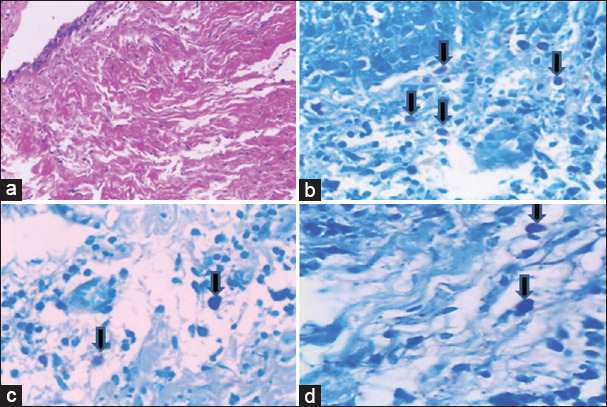
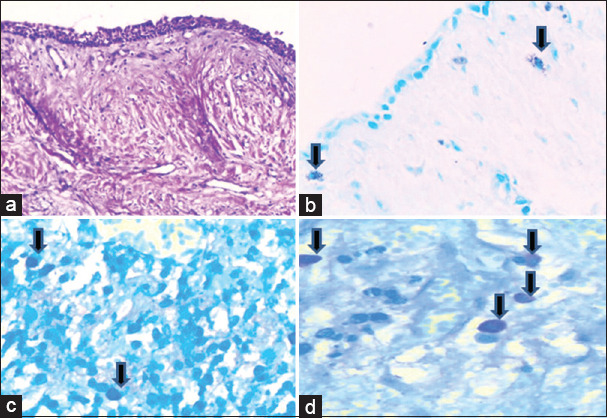
Introduction: Mast cells are large granular cells that arise from multipotent CD 34+ precursors in the bone marrow normally distributed throughout the connective tissues. Following activation of immunologic or nonimmunologic stimuli, mast cells release secretory granules which give the characteristic metachromatic appearance with toluidine blue stain. Release of numerous mediators on degranulation of mast cells plays an important role in the pathogenesis of odontogenic cysts.
Context: Odontogenic cysts, such as dentigerous cysts, arise due to the accumulation of fluid between the crown of an unerupted tooth and the reduced enamel epithelium. Dental follicles, which surround developing teeth, can also undergo cystic transformation. Mast cells activity might contribute to cyst expansion and bone resorption, highlighting their potential role in cystic pathology.
Aims: To study the presence of mast cells in the dental follicle and dentigerous cyst. To quantify the mast cells in the abovementioned subjects. To study the pattern of distribution of mast cell distribution in different zones of the study groups.
Settings and design: This was histopathological study conducted at the Department of Oral Pathology and Microbiology, Vydehi College of Dental Sciences and Research Centre, Bengaluru, between 2012 and 2015.
Methods and material: Our study was conducted in the Department of Oral Pathology and Microbiology at Vydehi College of Dental Sciences and Research Centre in the year 2012 to 2015. Histopathologically analyzed 30 cases each of dental follicle, and dentigerous cysts were taken and 4-5 micron sections were stained with toluidine blue. Counting of mast cells was done in three different zones which included subepithelial, intermediate, and deep zone. The results were tabulated and statistically analyzed.
Results: Both dental follicles and dentigerous cysts showed the presence of mast cells, and highest numbers of mast cells were seen in subepithelial zone followed by intermediate and deep zones. There was statistically significant relation in the number of mast cells in dentigerous cysts and dental follicle along subepithelial and intermediate zone with a P value of <0.05. In our study, we also found increased mast cell count in inflamed cases of dental follicle and dentigerous cyst compared with noninflamed cases with a P value of <0.01.
Conclusions: It is well known that mast cells play a role in the initiation of inflammation, and this inflammatory process may be associated with pericoronal follicle enlargement, a process that could result in cystic transformation of the follicle. Hence, regular radiographic follow-up is necessary especially for teeth with a maximum dental follicle width of 2-3 mm.
期刊介绍:
The journal of Oral and Maxillofacial Pathology [ISSN:print-(0973-029X, online-1998-393X)] is a tri-annual journal published on behalf of “The Indian Association of Oral and Maxillofacial Pathologists” (IAOMP). The publication of JOMFP was started in the year 1993. The journal publishes papers on a wide spectrum of topics associated with the scope of Oral and Maxillofacial Pathology, also, ensuring scientific merit and quality. It is a comprehensive reading material for the professionals who want to upgrade their diagnostic skills in Oral Diseases; allows exposure to newer topics and methods of research in the Oral-facial Tissues and Pathology. New features allow an open minded thinking and approach to various pathologies. It also encourages authors to showcase quality work done by them and to compile relevant cases which are diagnostically challenging. The Journal takes pride in maintaining the quality of articles and photomicrographs.

 求助内容:
求助内容: 应助结果提醒方式:


