Mustafa Kayabaşi, Seher Köksaldi, Neslihan Demirel, Ali Osman Saatci
{"title":"The Effect of Axial Length on Macular Vascular Density in Eyes with High Myopia.","authors":"Mustafa Kayabaşi, Seher Köksaldi, Neslihan Demirel, Ali Osman Saatci","doi":"10.22336/rjo.2025.15","DOIUrl":null,"url":null,"abstract":"<p><strong>Objective: </strong>To evaluate the relationship between optical coherence tomography angiography (OCTA) findings and axial length (AL) in eyes with high myopia.</p><p><strong>Materials and methods: </strong>A total of 122 eyes from 78 patients were included. Seventy-five eyes with an AL ranging between 26.00 and 27.49 mm comprised Group 1, and 47 with an AL of ≥ 27.50 mm comprised Group 2. Spectral-domain OCT was performed to measure the central macular thickness, subfoveal choroidal thickness (SCT) and swept-source OCTA was utilized to obtain the data on foveal avascular zone (FAZ) and vascular density (VD) values at the superficial and deep capillary plexuses (SCP and DCP), outer retina (OuR), and choriocapillaris (CC) segments.</p><p><strong>Results: </strong>While no significant differences were found in terms of the mean superficial-FAZ and deep-FAZ areas (<i>p</i>=0.284 and <i>p</i>=0.952, respectively), there were significant differences between the groups in terms of the mean foveal VD in the SCP (<i>p</i>=0.001), the mean total VD (<i>p</i>=0.045) and foveal VD in the DCP (<i>p</i><0.001), the mean foveal VD (<i>p</i>=0.019) and superior parafoveal VD in the OuR (<i>p</i>=0.008), the mean total (<i>p</i>=0.005), temporal parafoveal (<i>p</i>=0.034), inferior parafoveal (<i>p</i>=0.029), and nasal parafoveal VDs in the CC segments (<i>p</i>=0.005).</p><p><strong>Discussion: </strong>The findings of the present study highlight the complex interplay between axial elongation and retinal microvasculature, suggesting that factors beyond mechanical stretching may contribute to these alterations. The variability in the existing literature on this topic arises from inconsistencies in the definition of high myopia, the use of different OCTA devices, and heterogeneous study populations. By including eyes with myopic maculopathy and employing axial length-based classification, this study provides a broad representation of high myopia. However, its retrospective design, single-center setting, and monoracial cohort represent limitations. Future large-scale, prospective studies involving diverse populations are needed to elucidate further the pathophysiology of high myopia and its impact on retinal and choroidal microcirculation.</p><p><strong>Conclusions: </strong>Our study revealed that high-myopic eyes with longer ALs exhibited increased total VD in the DCP and increased foveal VD in the SCP, DCP, and OuR segments, while they showed decreased total VD and temporal, inferior, and nasal parafoveal VDs in the CC segment compared to high-myopic eyes with shorter ALs.</p>","PeriodicalId":94355,"journal":{"name":"Romanian journal of ophthalmology","volume":"69 1","pages":"88-100"},"PeriodicalIF":0.0000,"publicationDate":"2025-01-01","publicationTypes":"Journal Article","fieldsOfStudy":null,"isOpenAccess":false,"openAccessPdf":"https://www.ncbi.nlm.nih.gov/pmc/articles/PMC12049640/pdf/","citationCount":"0","resultStr":null,"platform":"Semanticscholar","paperid":null,"PeriodicalName":"Romanian journal of ophthalmology","FirstCategoryId":"1085","ListUrlMain":"https://doi.org/10.22336/rjo.2025.15","RegionNum":0,"RegionCategory":null,"ArticlePicture":[],"TitleCN":null,"AbstractTextCN":null,"PMCID":null,"EPubDate":"","PubModel":"","JCR":"","JCRName":"","Score":null,"Total":0}
引用次数: 0
Abstract
Objective: To evaluate the relationship between optical coherence tomography angiography (OCTA) findings and axial length (AL) in eyes with high myopia.
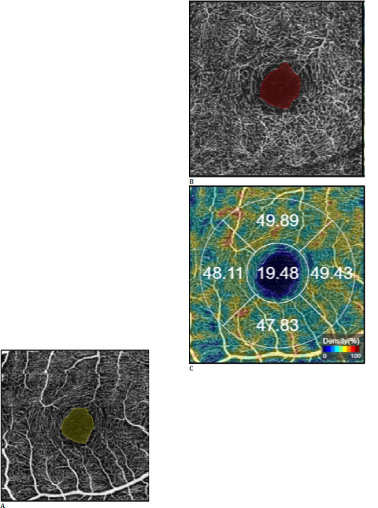
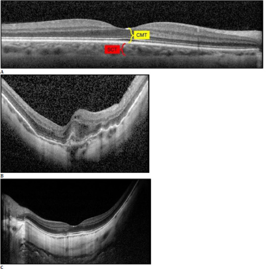
Materials and methods: A total of 122 eyes from 78 patients were included. Seventy-five eyes with an AL ranging between 26.00 and 27.49 mm comprised Group 1, and 47 with an AL of ≥ 27.50 mm comprised Group 2. Spectral-domain OCT was performed to measure the central macular thickness, subfoveal choroidal thickness (SCT) and swept-source OCTA was utilized to obtain the data on foveal avascular zone (FAZ) and vascular density (VD) values at the superficial and deep capillary plexuses (SCP and DCP), outer retina (OuR), and choriocapillaris (CC) segments.
Results: While no significant differences were found in terms of the mean superficial-FAZ and deep-FAZ areas (p=0.284 and p=0.952, respectively), there were significant differences between the groups in terms of the mean foveal VD in the SCP (p=0.001), the mean total VD (p=0.045) and foveal VD in the DCP (p<0.001), the mean foveal VD (p=0.019) and superior parafoveal VD in the OuR (p=0.008), the mean total (p=0.005), temporal parafoveal (p=0.034), inferior parafoveal (p=0.029), and nasal parafoveal VDs in the CC segments (p=0.005).
Discussion: The findings of the present study highlight the complex interplay between axial elongation and retinal microvasculature, suggesting that factors beyond mechanical stretching may contribute to these alterations. The variability in the existing literature on this topic arises from inconsistencies in the definition of high myopia, the use of different OCTA devices, and heterogeneous study populations. By including eyes with myopic maculopathy and employing axial length-based classification, this study provides a broad representation of high myopia. However, its retrospective design, single-center setting, and monoracial cohort represent limitations. Future large-scale, prospective studies involving diverse populations are needed to elucidate further the pathophysiology of high myopia and its impact on retinal and choroidal microcirculation.
Conclusions: Our study revealed that high-myopic eyes with longer ALs exhibited increased total VD in the DCP and increased foveal VD in the SCP, DCP, and OuR segments, while they showed decreased total VD and temporal, inferior, and nasal parafoveal VDs in the CC segment compared to high-myopic eyes with shorter ALs.

 求助内容:
求助内容: 应助结果提醒方式:


