{"title":"An extremely rare case of Rosai-Dorfman-Destombes disease in the spleen with secondary thrombocytopenia: a case report.","authors":"Xiqi Liu, Cheng Quan, Yu Wang","doi":"10.21037/acr-24-207","DOIUrl":null,"url":null,"abstract":"<p><strong>Background: </strong>Rosai-Dorfman-Destombes disease (RDD), also known as sinus histiocytosis with massive lymphadenopathy, is a rare, multisystemic histiocytic disorder. It can affect multiple organs, including bones, the brain, nasal cavities, and breasts. But, RDD in the spleen with secondary thrombocytopenia is extremely rare. This report aimed to show some new symptoms to help in the early diagnosis of this disease.</p><p><strong>Case description: </strong>A 68-year-old female patient presented with abdominal discomfort for over 2 months. Positron emission tomography-computed tomography (PET-CT) examination revealed multiple splenic lesions with no significant abnormalities elsewhere. The patient had a history of rheumatoid arthritis and diabetes. Physical examination showed no significant abnormalities. Blood tests upon admission revealed a platelet count of 39×10<sup>9</sup> cells/L. An elective laparoscopic splenectomy was performed in April 2024. Postoperative pathology and immunohistochemistry suggested RDD. Based on the lab reports and clinical manifestations, the patient was diagnosed with splenic primary RDD with secondary thrombocytopenia. The patient was followed up regularly, and the platelet level recovered to 222×10<sup>9</sup> cells/L 1-month post-surgery, confirming the cause of thrombocytopenia as secondary to splenic RDD. No significant abnormalities were found on abdominal CT 5 months post-surgery. Preoperative diagnosis of RDD remains challenging, especially for abdominal primary RDD, as percutaneous biopsy is difficult and imaging studies lack specific features, making the diagnosis still dependent on postoperative pathology and immunohistochemistry.</p><p><strong>Conclusions: </strong>This case indicates that in patients with multiple splenic space-occupying lesions and thrombocytopenia, particularly with a history of rheumatoid arthritis, the potential for this illness should be contemplated, even in the absence of conventional RDD lymph node symptoms.</p>","PeriodicalId":29752,"journal":{"name":"AME Case Reports","volume":"9 ","pages":"57"},"PeriodicalIF":0.7000,"publicationDate":"2025-03-11","publicationTypes":"Journal Article","fieldsOfStudy":null,"isOpenAccess":false,"openAccessPdf":"https://www.ncbi.nlm.nih.gov/pmc/articles/PMC12053440/pdf/","citationCount":"0","resultStr":null,"platform":"Semanticscholar","paperid":null,"PeriodicalName":"AME Case Reports","FirstCategoryId":"1085","ListUrlMain":"https://doi.org/10.21037/acr-24-207","RegionNum":0,"RegionCategory":null,"ArticlePicture":[],"TitleCN":null,"AbstractTextCN":null,"PMCID":null,"EPubDate":"2025/1/1 0:00:00","PubModel":"eCollection","JCR":"Q3","JCRName":"MEDICINE, GENERAL & INTERNAL","Score":null,"Total":0}
引用次数: 0
Abstract
Background: Rosai-Dorfman-Destombes disease (RDD), also known as sinus histiocytosis with massive lymphadenopathy, is a rare, multisystemic histiocytic disorder. It can affect multiple organs, including bones, the brain, nasal cavities, and breasts. But, RDD in the spleen with secondary thrombocytopenia is extremely rare. This report aimed to show some new symptoms to help in the early diagnosis of this disease.
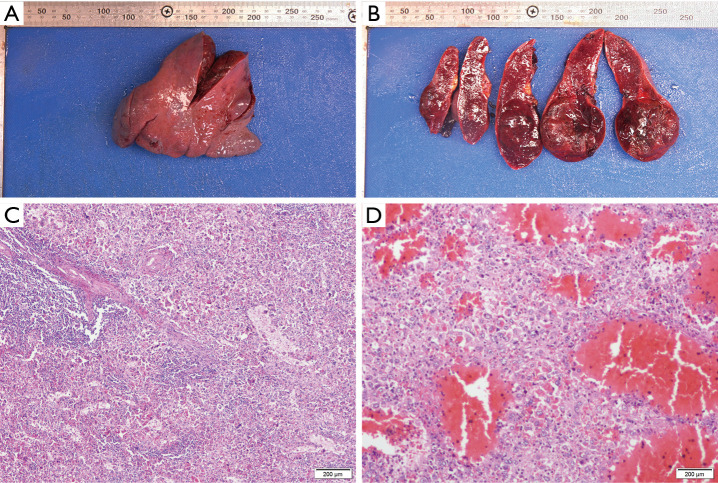
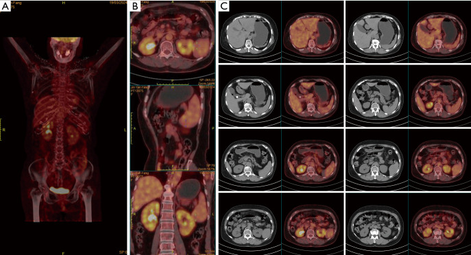
Case description: A 68-year-old female patient presented with abdominal discomfort for over 2 months. Positron emission tomography-computed tomography (PET-CT) examination revealed multiple splenic lesions with no significant abnormalities elsewhere. The patient had a history of rheumatoid arthritis and diabetes. Physical examination showed no significant abnormalities. Blood tests upon admission revealed a platelet count of 39×109 cells/L. An elective laparoscopic splenectomy was performed in April 2024. Postoperative pathology and immunohistochemistry suggested RDD. Based on the lab reports and clinical manifestations, the patient was diagnosed with splenic primary RDD with secondary thrombocytopenia. The patient was followed up regularly, and the platelet level recovered to 222×109 cells/L 1-month post-surgery, confirming the cause of thrombocytopenia as secondary to splenic RDD. No significant abnormalities were found on abdominal CT 5 months post-surgery. Preoperative diagnosis of RDD remains challenging, especially for abdominal primary RDD, as percutaneous biopsy is difficult and imaging studies lack specific features, making the diagnosis still dependent on postoperative pathology and immunohistochemistry.
Conclusions: This case indicates that in patients with multiple splenic space-occupying lesions and thrombocytopenia, particularly with a history of rheumatoid arthritis, the potential for this illness should be contemplated, even in the absence of conventional RDD lymph node symptoms.

 求助内容:
求助内容: 应助结果提醒方式:


