{"title":"Spectral computed tomography parameters of primary tumors and lymph nodes for predicting tumor deposits in colorectal cancer.","authors":"Yi-Fan Lai, Zhao-Ming Liang, Jing-Fang Li, Jia-Ying Zhang, Ding-Hua Xu, Hai-Yang Dai","doi":"10.4329/wjr.v17.i4.103359","DOIUrl":null,"url":null,"abstract":"<p><strong>Background: </strong>Tumor deposits (TDs) are an independent predictor of poor prognosis in colorectal cancer (CRC) patients. Enhanced follow-up and treatment monitoring for TD+ patients may improve survival rates and quality of life. However, the detection of TDs relies primarily on postoperative pathological examination, which may have a low detection rate due to sampling limitations.</p><p><strong>Aim: </strong>To evaluate the spectral computed tomography (CT) parameters of primary tumors and the largest regional lymph nodes (LNs), to determine their value in predicting TDs in CRC.</p><p><strong>Methods: </strong>A retrospective analysis was conducted which included 121 patients with CRC whose complete spectral CT data were available. Patients were divided into the TDs+ group and the TDs- group on the basis of their pathological results. Spectral CT parameters of the primary CRC lesion and the largest regional LNs were measured, including the normalized iodine concentration (NIC) in both the arterial and venous phases, and the LN-to-primary tumor ratio was calculated. Statistical methods were used to evaluate the diagnostic efficacy of each spectral parameter.</p><p><strong>Results: </strong>Among the 121 CRC patients, 33 (27.2%) were confirmed to be TDs+. The risk of TDs positivity was greater in patients with positive LN metastasis, higher N stage and elevated carcinoembryonic antigen and cancer antigen 19-9 levels. The NIC (LNs in both the arterial and venous phases), NIC (primary tumors in the venous phase), and the LN-to-primary tumor ratio in both the arterial and venous phases were associated with TDs (<i>P</i> < 0.05). In multivariate logistic regression analysis, the arterial phase LN-to-primary tumor ratio was identified as an independent predictor of TDs, demonstrating the highest diagnostic performance (area under the curve: 0.812, sensitivity: 0.879, specificity: 0.648, cutoff value: 1.145).</p><p><strong>Conclusion: </strong>The spectral CT parameters of the primary colorectal tumor and the largest regional LNs, especially the LN-to-primary tumor ratio, have significant clinical value in predicting TDs in CRC.</p>","PeriodicalId":23819,"journal":{"name":"World journal of radiology","volume":"17 4","pages":"103359"},"PeriodicalIF":1.5000,"publicationDate":"2025-04-28","publicationTypes":"Journal Article","fieldsOfStudy":null,"isOpenAccess":false,"openAccessPdf":"https://www.ncbi.nlm.nih.gov/pmc/articles/PMC12038403/pdf/","citationCount":"0","resultStr":null,"platform":"Semanticscholar","paperid":null,"PeriodicalName":"World journal of radiology","FirstCategoryId":"1085","ListUrlMain":"https://doi.org/10.4329/wjr.v17.i4.103359","RegionNum":0,"RegionCategory":null,"ArticlePicture":[],"TitleCN":null,"AbstractTextCN":null,"PMCID":null,"EPubDate":"","PubModel":"","JCR":"Q3","JCRName":"RADIOLOGY, NUCLEAR MEDICINE & MEDICAL IMAGING","Score":null,"Total":0}
引用次数: 0
Abstract
Background: Tumor deposits (TDs) are an independent predictor of poor prognosis in colorectal cancer (CRC) patients. Enhanced follow-up and treatment monitoring for TD+ patients may improve survival rates and quality of life. However, the detection of TDs relies primarily on postoperative pathological examination, which may have a low detection rate due to sampling limitations.
Aim: To evaluate the spectral computed tomography (CT) parameters of primary tumors and the largest regional lymph nodes (LNs), to determine their value in predicting TDs in CRC.
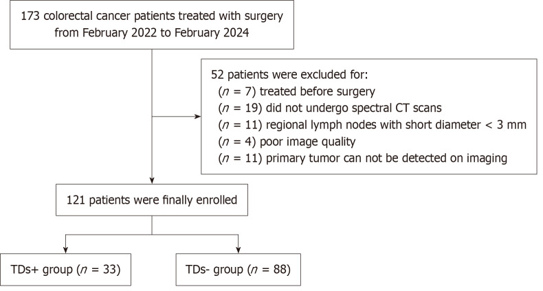
Methods: A retrospective analysis was conducted which included 121 patients with CRC whose complete spectral CT data were available. Patients were divided into the TDs+ group and the TDs- group on the basis of their pathological results. Spectral CT parameters of the primary CRC lesion and the largest regional LNs were measured, including the normalized iodine concentration (NIC) in both the arterial and venous phases, and the LN-to-primary tumor ratio was calculated. Statistical methods were used to evaluate the diagnostic efficacy of each spectral parameter.
Results: Among the 121 CRC patients, 33 (27.2%) were confirmed to be TDs+. The risk of TDs positivity was greater in patients with positive LN metastasis, higher N stage and elevated carcinoembryonic antigen and cancer antigen 19-9 levels. The NIC (LNs in both the arterial and venous phases), NIC (primary tumors in the venous phase), and the LN-to-primary tumor ratio in both the arterial and venous phases were associated with TDs (P < 0.05). In multivariate logistic regression analysis, the arterial phase LN-to-primary tumor ratio was identified as an independent predictor of TDs, demonstrating the highest diagnostic performance (area under the curve: 0.812, sensitivity: 0.879, specificity: 0.648, cutoff value: 1.145).
Conclusion: The spectral CT parameters of the primary colorectal tumor and the largest regional LNs, especially the LN-to-primary tumor ratio, have significant clinical value in predicting TDs in CRC.

 求助内容:
求助内容: 应助结果提醒方式:


