{"title":"Enhanced Electroacoustic Tomography with Supervised Learning for Real-time Electroporation Monitoring.","authors":"Zhuoran Jiang, Yifei Xu, Leshan Sun, Shreyas Srinivasan, Q Jackie Wu, Liangzhong Xiang, Lei Ren","doi":"10.1002/pro6.1242","DOIUrl":null,"url":null,"abstract":"<p><strong>Background: </strong>Nanosecond pulsed electric fields (nsPEF)-based electroporation is a new therapy modality potentially synergized with radiation therapy to improve treatment outcomes. To verify its treatment accuracy intraoperatively, electroacoustic tomography (EAT) has been developed to monitor in-vivo electric energy deposition by detecting ultrasound signals generated by nsPEFs in real-time. However, utility of EAT is limited by image distortions due to the limited-angle view of ultrasound transducers.</p><p><strong>Methods: </strong>This study proposed a supervised learning-based workflow to address the ill-conditioning in EAT reconstruction. Electroacoustic signals were detected by a linear array and initially reconstructed into EAT images, which were then fed into a deep learning model for distortion correction. In this study, 56 distinct electroacoustic data sets from nsPEFs of different intensities and geometries were collected experimentally, avoiding simulation-to-real-world variations. Forty-six data were used for model training and 10 for testing. The model was trained using supervised learning, enabled by a custom rotating platform to acquire paired full-view and single-view signals for the same electric field.</p><p><strong>Results: </strong>The proposed method considerably improved the image quality of linear array-based EAT, generating pressure maps with accurate and clear structures. Quantitatively, the enhanced single-view images achieved a low-intensity error (RMSE: 0.018), high signal-to-noise ratio (PSNR: 35.15), and high structural similarity (SSIM: 0.942) compared to the reference full-view images.</p><p><strong>Conclusions: </strong>This study represented a pioneering stride in achieving high-quality EAT using a single linear array in an experimental environment, which improves EAT's utility in real-time monitoring for nsPEF-based electroporation therapy.</p>","PeriodicalId":32406,"journal":{"name":"Precision Radiation Oncology","volume":"8 3","pages":"110-118"},"PeriodicalIF":2.1000,"publicationDate":"2024-09-22","publicationTypes":"Journal Article","fieldsOfStudy":null,"isOpenAccess":false,"openAccessPdf":"https://www.ncbi.nlm.nih.gov/pmc/articles/PMC11935180/pdf/","citationCount":"0","resultStr":null,"platform":"Semanticscholar","paperid":null,"PeriodicalName":"Precision Radiation Oncology","FirstCategoryId":"1085","ListUrlMain":"https://doi.org/10.1002/pro6.1242","RegionNum":0,"RegionCategory":null,"ArticlePicture":[],"TitleCN":null,"AbstractTextCN":null,"PMCID":null,"EPubDate":"2024/9/1 0:00:00","PubModel":"eCollection","JCR":"Q4","JCRName":"Medicine","Score":null,"Total":0}
引用次数: 0
Abstract
Background: Nanosecond pulsed electric fields (nsPEF)-based electroporation is a new therapy modality potentially synergized with radiation therapy to improve treatment outcomes. To verify its treatment accuracy intraoperatively, electroacoustic tomography (EAT) has been developed to monitor in-vivo electric energy deposition by detecting ultrasound signals generated by nsPEFs in real-time. However, utility of EAT is limited by image distortions due to the limited-angle view of ultrasound transducers.
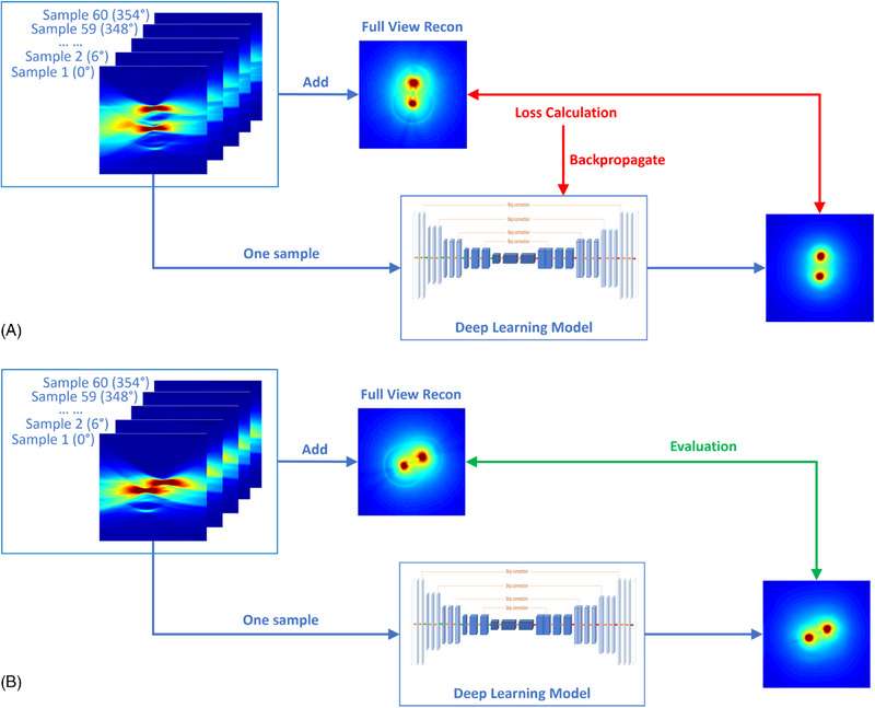
Methods: This study proposed a supervised learning-based workflow to address the ill-conditioning in EAT reconstruction. Electroacoustic signals were detected by a linear array and initially reconstructed into EAT images, which were then fed into a deep learning model for distortion correction. In this study, 56 distinct electroacoustic data sets from nsPEFs of different intensities and geometries were collected experimentally, avoiding simulation-to-real-world variations. Forty-six data were used for model training and 10 for testing. The model was trained using supervised learning, enabled by a custom rotating platform to acquire paired full-view and single-view signals for the same electric field.
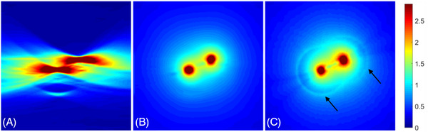
Results: The proposed method considerably improved the image quality of linear array-based EAT, generating pressure maps with accurate and clear structures. Quantitatively, the enhanced single-view images achieved a low-intensity error (RMSE: 0.018), high signal-to-noise ratio (PSNR: 35.15), and high structural similarity (SSIM: 0.942) compared to the reference full-view images.
Conclusions: This study represented a pioneering stride in achieving high-quality EAT using a single linear array in an experimental environment, which improves EAT's utility in real-time monitoring for nsPEF-based electroporation therapy.

 求助内容:
求助内容: 应助结果提醒方式:


