{"title":"Acute anterior uveitis after photorefractive keratectomy: Demographic profile and clinical characteristics.","authors":"Ashish Markan, Shivani Chabbra, Mohammed Ibrahime Asif, Rahil Chaudhary, Manasi Tripathi","doi":"10.22336/rjo.2025.09","DOIUrl":null,"url":null,"abstract":"<p><strong>Objective: </strong>To report demographic profile and clinical characteristics of acute anterior uveitis (AAU) after Photorefractive Keratectomy (PRK).</p><p><strong>Materials and methods: </strong>This retrospective study reviewed records of all patients who underwent PRK to correct ametropia between July 2021 and June 2023. Patients who developed postoperative AAU were included for evaluation. Demographic details, preoperative ocular examination, intraoperative details, postoperative examination, time to onset of AAU, grading of cells, flare and pigments, time to heal, and final visual outcome were analyzed.</p><p><strong>Results: </strong>Records of 390 patients who underwent PRK during the study period were reviewed. Of these, 16 (29 eyes, b/l:13 patients, u/l: 3 patients) presented with AAU following PRK. The mean age of patients was 27.43 + 4.53 years (range 22-32 years), with a mean spherical equivalent of -3.18 + 2.16 (range -2 to -7 D). Mean ablation depth was 55.13 + 28.10 mm (range 27-105 mm), and mean duration of excimer laser ablation was 11.27 + 8.31 seconds (range 3-36 seconds). The mean onset time of AAU post-surgery was 27.8 + 10.9 days (range 7-47 days). Most eyes (75.86%, n=22) had a moderate-intense grade of inflammation, while 82.75% (n=24) of the eyes had significant pigment dispersion. The mean healing time was 57.43 + 27.87 days (33-106 days). The median follow-up duration was 12 months (range 6-18 months). The incidence of AAU post-PRK in our study was 4.1%.</p><p><strong>Discussion: </strong>This study's incidence of post-PRK acute anterior uveitis (AAU) was 4.1%, aligning with previous reports of rare but significant inflammatory responses following PRK. The mean onset at 27.8 days suggested a delayed immune-mediated reaction rather than an immediate post-surgical response. While most cases had mild-to-moderate inflammation, a subset experienced severe reactions and ocular hypertension, reinforcing the need for close monitoring. The absence of systemic associations and posterior segment involvement suggested the role of a localized immune response. Despite the prolonged resolution time, all eyes achieved a final CDVA of 0 LogMAR, indicating favorable long-term outcomes with timely intervention and management.</p><p><strong>Conclusion: </strong>Anterior uveitis following PRK is infrequent. While it presents with marked anterior chamber reaction and pigment dispersion, the inflammation is often well-controlled with topical steroids. It does not affect the final visual outcome.</p>","PeriodicalId":94355,"journal":{"name":"Romanian journal of ophthalmology","volume":"69 1","pages":"48-52"},"PeriodicalIF":0.0000,"publicationDate":"2025-01-01","publicationTypes":"Journal Article","fieldsOfStudy":null,"isOpenAccess":false,"openAccessPdf":"https://www.ncbi.nlm.nih.gov/pmc/articles/PMC12049649/pdf/","citationCount":"0","resultStr":null,"platform":"Semanticscholar","paperid":null,"PeriodicalName":"Romanian journal of ophthalmology","FirstCategoryId":"1085","ListUrlMain":"https://doi.org/10.22336/rjo.2025.09","RegionNum":0,"RegionCategory":null,"ArticlePicture":[],"TitleCN":null,"AbstractTextCN":null,"PMCID":null,"EPubDate":"","PubModel":"","JCR":"","JCRName":"","Score":null,"Total":0}
引用次数: 0
Abstract
Objective: To report demographic profile and clinical characteristics of acute anterior uveitis (AAU) after Photorefractive Keratectomy (PRK).
Materials and methods: This retrospective study reviewed records of all patients who underwent PRK to correct ametropia between July 2021 and June 2023. Patients who developed postoperative AAU were included for evaluation. Demographic details, preoperative ocular examination, intraoperative details, postoperative examination, time to onset of AAU, grading of cells, flare and pigments, time to heal, and final visual outcome were analyzed.
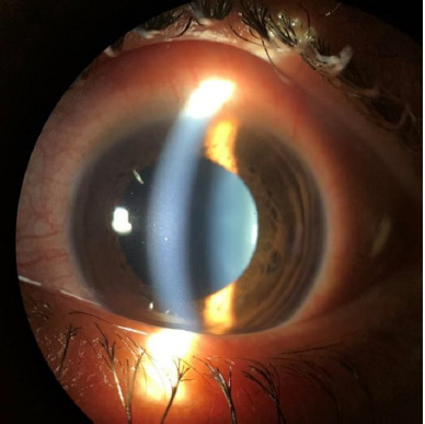
Results: Records of 390 patients who underwent PRK during the study period were reviewed. Of these, 16 (29 eyes, b/l:13 patients, u/l: 3 patients) presented with AAU following PRK. The mean age of patients was 27.43 + 4.53 years (range 22-32 years), with a mean spherical equivalent of -3.18 + 2.16 (range -2 to -7 D). Mean ablation depth was 55.13 + 28.10 mm (range 27-105 mm), and mean duration of excimer laser ablation was 11.27 + 8.31 seconds (range 3-36 seconds). The mean onset time of AAU post-surgery was 27.8 + 10.9 days (range 7-47 days). Most eyes (75.86%, n=22) had a moderate-intense grade of inflammation, while 82.75% (n=24) of the eyes had significant pigment dispersion. The mean healing time was 57.43 + 27.87 days (33-106 days). The median follow-up duration was 12 months (range 6-18 months). The incidence of AAU post-PRK in our study was 4.1%.
Discussion: This study's incidence of post-PRK acute anterior uveitis (AAU) was 4.1%, aligning with previous reports of rare but significant inflammatory responses following PRK. The mean onset at 27.8 days suggested a delayed immune-mediated reaction rather than an immediate post-surgical response. While most cases had mild-to-moderate inflammation, a subset experienced severe reactions and ocular hypertension, reinforcing the need for close monitoring. The absence of systemic associations and posterior segment involvement suggested the role of a localized immune response. Despite the prolonged resolution time, all eyes achieved a final CDVA of 0 LogMAR, indicating favorable long-term outcomes with timely intervention and management.
Conclusion: Anterior uveitis following PRK is infrequent. While it presents with marked anterior chamber reaction and pigment dispersion, the inflammation is often well-controlled with topical steroids. It does not affect the final visual outcome.

 求助内容:
求助内容: 应助结果提醒方式:


