Yubo Guo, Lu Lin, Keting Xu, Shihai Zhao, Gan Sun, Yuyan Chen, Ke Xue, Yuxin Yang, Shuo Chen, Yan Zhang, Yanjie Zhu, Yining Wang
{"title":"Myocardial native T1 and extracellular volume measurements at 5T: Feasibility study and initial experience.","authors":"Yubo Guo, Lu Lin, Keting Xu, Shihai Zhao, Gan Sun, Yuyan Chen, Ke Xue, Yuxin Yang, Shuo Chen, Yan Zhang, Yanjie Zhu, Yining Wang","doi":"10.1016/j.jocmr.2025.101896","DOIUrl":null,"url":null,"abstract":"<p><strong>Background: </strong>T1 mapping is a robust and highly reproducible technique for quantitative assessment of cardiomyopathy. The aim of this study is to investigate the feasibility of 5T myocardial T1 mapping and to establish preliminary reference values for myocardial T1 at 5T.</p><p><strong>Methods: </strong>Twenty-eight healthy volunteers (median age, 42 [interquartile range (IQR): 29-54] years; 14 male) and 11 patients (median age, 44 [IQR: 34-51] years; 7 male) underwent cardiovascular magnetic resonance at 5T. T1 mapping was acquired using a motion-corrected modified Look-Locker inversion recovery sequence [5(3)3 scheme for pre-contrast, (4(1)3(1)2) scheme for post-contrast] in three short-axis planes (base, middle, and apex). T1 values were quantified per segment, per slice, and globally.</p><p><strong>Results: </strong>Healthy volunteers had a mean global native T1 value of 1506.2 ± 41.7 ms, with T1 values decreasing progressively from the base to the apex slice (P = 0.08). Significantly higher T1 values were revealed in the septum compared to the non-septal myocardium (1540.1 ± 69.3 vs 1477.6 ± 93.7, P < 0.001). No statistically significant gender- and age-related differences were observed in native T1 values (both, P > 0.05). Within the spectrum of cardiac pathologies analyzed in this study, patients exhibited higher native T1 values (1638.7 ± 108.6 ms vs 1506.2 ± 41.7 ms, P < 0.001) and higher extracellular volume fraction (37.5% ± 5.5% vs 29.5% ± 2.1%, P = 0.074) compared to healthy volunteers, late gadolinium enhancement (LGE)-positive segments exhibited significantly higher T1 values than LGE-negative segments (1685.2 ± 144.1 vs 1582.6 ± 88.7, P < 0.001). There was excellent intra-scanner test-retest, intra-observer, and inter-observer reproducibility for measurement of native T1.</p><p><strong>Conclusion: </strong>The present study demonstrated the feasibility of T1 mapping quantification at 5T and presented mean native T1 values in healthy human myocardium at this field strength, which can be used as reference values specific for this magnetic resonance setting.</p>","PeriodicalId":15221,"journal":{"name":"Journal of Cardiovascular Magnetic Resonance","volume":"27 1","pages":"101896"},"PeriodicalIF":6.1000,"publicationDate":"2025-01-01","publicationTypes":"Journal Article","fieldsOfStudy":null,"isOpenAccess":false,"openAccessPdf":"https://www.ncbi.nlm.nih.gov/pmc/articles/PMC12182820/pdf/","citationCount":"0","resultStr":null,"platform":"Semanticscholar","paperid":null,"PeriodicalName":"Journal of Cardiovascular Magnetic Resonance","FirstCategoryId":"3","ListUrlMain":"https://doi.org/10.1016/j.jocmr.2025.101896","RegionNum":1,"RegionCategory":"医学","ArticlePicture":[],"TitleCN":null,"AbstractTextCN":null,"PMCID":null,"EPubDate":"2025/4/21 0:00:00","PubModel":"Epub","JCR":"Q1","JCRName":"CARDIAC & CARDIOVASCULAR SYSTEMS","Score":null,"Total":0}
引用次数: 0
Abstract
Background: T1 mapping is a robust and highly reproducible technique for quantitative assessment of cardiomyopathy. The aim of this study is to investigate the feasibility of 5T myocardial T1 mapping and to establish preliminary reference values for myocardial T1 at 5T.
Methods: Twenty-eight healthy volunteers (median age, 42 [interquartile range (IQR): 29-54] years; 14 male) and 11 patients (median age, 44 [IQR: 34-51] years; 7 male) underwent cardiovascular magnetic resonance at 5T. T1 mapping was acquired using a motion-corrected modified Look-Locker inversion recovery sequence [5(3)3 scheme for pre-contrast, (4(1)3(1)2) scheme for post-contrast] in three short-axis planes (base, middle, and apex). T1 values were quantified per segment, per slice, and globally.
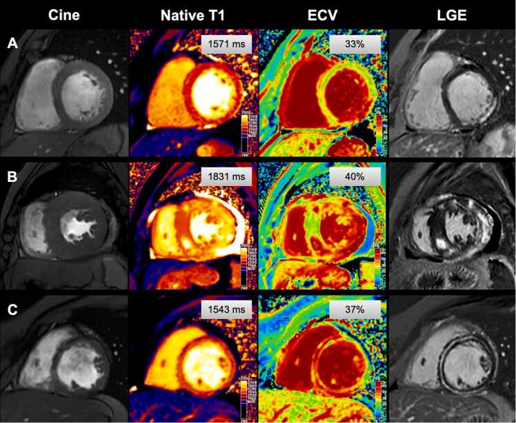
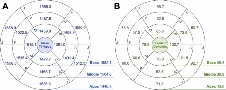
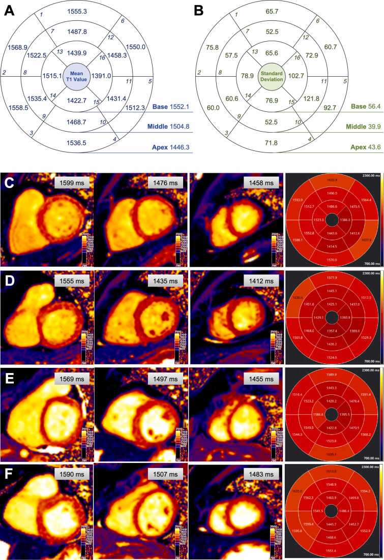
Results: Healthy volunteers had a mean global native T1 value of 1506.2 ± 41.7 ms, with T1 values decreasing progressively from the base to the apex slice (P = 0.08). Significantly higher T1 values were revealed in the septum compared to the non-septal myocardium (1540.1 ± 69.3 vs 1477.6 ± 93.7, P < 0.001). No statistically significant gender- and age-related differences were observed in native T1 values (both, P > 0.05). Within the spectrum of cardiac pathologies analyzed in this study, patients exhibited higher native T1 values (1638.7 ± 108.6 ms vs 1506.2 ± 41.7 ms, P < 0.001) and higher extracellular volume fraction (37.5% ± 5.5% vs 29.5% ± 2.1%, P = 0.074) compared to healthy volunteers, late gadolinium enhancement (LGE)-positive segments exhibited significantly higher T1 values than LGE-negative segments (1685.2 ± 144.1 vs 1582.6 ± 88.7, P < 0.001). There was excellent intra-scanner test-retest, intra-observer, and inter-observer reproducibility for measurement of native T1.
Conclusion: The present study demonstrated the feasibility of T1 mapping quantification at 5T and presented mean native T1 values in healthy human myocardium at this field strength, which can be used as reference values specific for this magnetic resonance setting.
背景:T1定位是一种可靠的、高度可重复的心肌病定量评估技术。本研究旨在探讨5T心肌T1制图的可行性,并建立5T心肌T1的初步参考值。方法:28名健康志愿者(年龄中位数42岁[四分位数间距29-54]岁;男性14例,患者11例(中位年龄44岁[IQR: 34-51]岁;7名男性)在5T行心血管磁共振。使用运动校正后的改进Look-Locker反演恢复序列[对比前采用5(3)3方案,对比后采用(4(1)3(1)2)方案]在三个短轴平面(基底、中间和顶点)获得T1映射。T1值被量化为每段、每片和全局。结果:健康志愿者的平均T1值为1506.2±41.7 ms, T1值从基底片到尖端片逐渐降低(P = 0.08)。与非间隔心肌相比,间隔心肌T1值明显高于间隔心肌(1540.1±69.3 vs 1477.6±93.7,P < 0.001)。原生T1值在性别和年龄方面无统计学差异(均P < 0.05)。在本研究分析的心脏病理谱中,与健康志愿者相比,患者具有更高的原生T1值(1638.7±108.6 ms vs 1506.2±41.7 ms, P < 0.001)和更高的细胞外体积分数(37.5%±5.5% vs 29.5%±2.1%,P = 0.074),晚期钆增强(LGE)阳性段的T1值明显高于LGE阴性段(1685.2±144.1 vs 1582.6±88.7,P < 0.001)。本机T1测量具有出色的扫描内测试-重测、观察者内和观察者间再现性。结论:本研究证明了5T时T1作图量化的可行性,并给出了该场强下健康心肌的平均天然T1值,可作为该磁共振设置的参考值。
期刊介绍:
Journal of Cardiovascular Magnetic Resonance (JCMR) publishes high-quality articles on all aspects of basic, translational and clinical research on the design, development, manufacture, and evaluation of cardiovascular magnetic resonance (CMR) methods applied to the cardiovascular system. Topical areas include, but are not limited to:
New applications of magnetic resonance to improve the diagnostic strategies, risk stratification, characterization and management of diseases affecting the cardiovascular system.
New methods to enhance or accelerate image acquisition and data analysis.
Results of multicenter, or larger single-center studies that provide insight into the utility of CMR.
Basic biological perceptions derived by CMR methods.

 求助内容:
求助内容: 应助结果提醒方式:


