Beth Whittington, Viswan Thiagarajah, Evangelos Tzolos, Jakub Kaczynski, Caelan Taggart, Alex Vesey, Damini Dey, Rachael O Forsythe, Andrew Tambyraja, Edwin J R van Beek, Marc R Dweck, David E Newby, Michelle C Williams
{"title":"Quantification of carotid artery plaque and peri-vascular adipose tissue attenuation on computed tomography.","authors":"Beth Whittington, Viswan Thiagarajah, Evangelos Tzolos, Jakub Kaczynski, Caelan Taggart, Alex Vesey, Damini Dey, Rachael O Forsythe, Andrew Tambyraja, Edwin J R van Beek, Marc R Dweck, David E Newby, Michelle C Williams","doi":"10.1093/ehjimp/qyaf040","DOIUrl":null,"url":null,"abstract":"<p><strong>Aims: </strong>Quantitative assessment of carotid artery plaque on computed tomography (CT) may identify high-risk phenotypes associated with culprit lesions and subsequent ischaemic stroke or transient ischaemic attack.</p><p><strong>Methods and results: </strong>Carotid CT angiography was performed in 48 patients with acute ischaemic stroke or transient ischaemic attack within 21 days. Quantitative plaque assessment was performed in the proximal 6 cm of the internal and external carotid artery, distal 6 cm of the common carotid artery, and residual common carotid artery. Semi-automated quantification included assessment of non-calcified, calcified, low-attenuation, and total plaque, area and diameter stenosis, and peri-vascular adipose tissue attenuation. In 48 patients (mean age 71 ± 11 years, 67% male), 96 vessels were assessed with 30 (31%) identified as culprit vessels. Culprit internal carotid arteries had greater area [83 (65, 94) vs. 64 (55, 77)%] and diameter [56 (39, 74) vs. 32 (21, 48)%] stenosis and more non-calcified [563 (413, 965) vs. 428 (283 649) mm<sup>3</sup>, <i>P</i> = 0.04], low-attenuation [33.7 (6.9, 72.4) vs. 16.3 (3.35, 54.3) mm<sup>3</sup>, <i>P</i> = 0.01], and total [699 (455, 1057) vs. 492 (311, 809), <i>P</i> = 0.04] plaque. There was no difference in calcified plaque or peri-vascular adipose tissue attenuation between culprit and non-culprit internal carotid arteries. There were no differences in quantitative plaque or peri-vascular adipose tissue attenuation in the external carotid artery or common carotid artery.</p><p><strong>Conclusion: </strong>Carotid atherosclerotic plaque characteristics are the principal features associated with culprit plaques with little or no demonstrable relationship with calcified plaque or increased peri-vascular adipose tissue attenuation.</p>","PeriodicalId":94317,"journal":{"name":"European heart journal. Imaging methods and practice","volume":"3 1","pages":"qyaf040"},"PeriodicalIF":0.0000,"publicationDate":"2025-04-08","publicationTypes":"Journal Article","fieldsOfStudy":null,"isOpenAccess":false,"openAccessPdf":"https://www.ncbi.nlm.nih.gov/pmc/articles/PMC12023745/pdf/","citationCount":"0","resultStr":null,"platform":"Semanticscholar","paperid":null,"PeriodicalName":"European heart journal. Imaging methods and practice","FirstCategoryId":"1085","ListUrlMain":"https://doi.org/10.1093/ehjimp/qyaf040","RegionNum":0,"RegionCategory":null,"ArticlePicture":[],"TitleCN":null,"AbstractTextCN":null,"PMCID":null,"EPubDate":"2025/1/1 0:00:00","PubModel":"eCollection","JCR":"","JCRName":"","Score":null,"Total":0}
引用次数: 0
Abstract
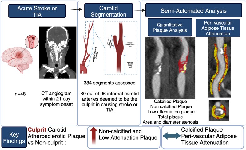
Aims: Quantitative assessment of carotid artery plaque on computed tomography (CT) may identify high-risk phenotypes associated with culprit lesions and subsequent ischaemic stroke or transient ischaemic attack.
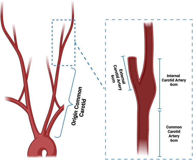
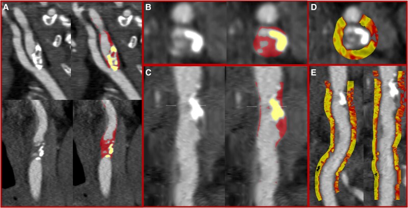
Methods and results: Carotid CT angiography was performed in 48 patients with acute ischaemic stroke or transient ischaemic attack within 21 days. Quantitative plaque assessment was performed in the proximal 6 cm of the internal and external carotid artery, distal 6 cm of the common carotid artery, and residual common carotid artery. Semi-automated quantification included assessment of non-calcified, calcified, low-attenuation, and total plaque, area and diameter stenosis, and peri-vascular adipose tissue attenuation. In 48 patients (mean age 71 ± 11 years, 67% male), 96 vessels were assessed with 30 (31%) identified as culprit vessels. Culprit internal carotid arteries had greater area [83 (65, 94) vs. 64 (55, 77)%] and diameter [56 (39, 74) vs. 32 (21, 48)%] stenosis and more non-calcified [563 (413, 965) vs. 428 (283 649) mm3, P = 0.04], low-attenuation [33.7 (6.9, 72.4) vs. 16.3 (3.35, 54.3) mm3, P = 0.01], and total [699 (455, 1057) vs. 492 (311, 809), P = 0.04] plaque. There was no difference in calcified plaque or peri-vascular adipose tissue attenuation between culprit and non-culprit internal carotid arteries. There were no differences in quantitative plaque or peri-vascular adipose tissue attenuation in the external carotid artery or common carotid artery.
Conclusion: Carotid atherosclerotic plaque characteristics are the principal features associated with culprit plaques with little or no demonstrable relationship with calcified plaque or increased peri-vascular adipose tissue attenuation.

 求助内容:
求助内容: 应助结果提醒方式:


