Ece Ozal, Sadik Altan Ozal, Riza Serttas, Suat Erdogan
{"title":"Unraveling the Role of Midkine in Proliferative Diabetic Retinopathy: Implications from Hypoxia-Induced Angiogenesis.","authors":"Ece Ozal, Sadik Altan Ozal, Riza Serttas, Suat Erdogan","doi":"10.14744/SEMB.2025.29964","DOIUrl":null,"url":null,"abstract":"<p><strong>Objectives: </strong>This study aimed to compare the expression of midkine (MK) in the vitreous of patients with proliferative diabetic retinopathy (PDR) and non-diabetic individuals, elucidating its potential role in the pathogenesis of the disease.</p><p><strong>Methods: </strong>This prospective cross-sectional study included three groups of patients who underwent pars plana vitrectomy (PPV) surgery. The first group (control) consisted of patients who underwent PPV for epiretinal membrane and macular hole and did not have diabetes mellitus (DM). The second group included patients who underwent PPV for vitreous hemorrhage (VH) and tractional retinal detachment (TRD) secondary to PDR without prior anti-VEGF treatment (No preoperative anti-VEGF application: NPa-VEGF). The third group comprised patients who underwent PPV for VH and TRD secondary to PDR and received a preoperative anti-VEGF injection one week before surgery (preoperative anti-VEGF application: Pa-VEGF). Vitreous samples were collected intraoperatively, and the concentrations of MK, interleukin (IL)-6, and IL-8 were measured using specific Enzyme-Linked Immunosorbent Assay (ELISA) kits.</p><p><strong>Results: </strong>The study included a total of 49 eyes from 49 patients undergoing PPV. The concentrations of IL-6 and IL-8 in vitreous samples from the NPa-VEGF group (n=15) and the Pa-VEGF group (n=14) were not significantly different compared to the control group (n=20) (p>0.05). However, the vitreous fluid of patients in the NPa-VEGF group exhibited significantly higher MK concentrations compared to the control group (p<0.007). Similarly, MK concentrations were significantly elevated in the Pa-VEGF group compared to the control group (p<0.046). No significant difference in MK levels was detected between the NPa-VEGF and Pa-VEGF groups (p>0.05).</p><p><strong>Conclusion: </strong>These findings suggest that increased MK expression in the vitreous may be associated with the pathogenesis of PDR. Further studies are warranted to elucidate the precise mechanisms underlying this association and to explore the potential of MK as a therapeutic target for PDR management.</p>","PeriodicalId":42218,"journal":{"name":"Medical Bulletin of Sisli Etfal Hospital","volume":"59 1","pages":"76-82"},"PeriodicalIF":0.9000,"publicationDate":"2025-03-18","publicationTypes":"Journal Article","fieldsOfStudy":null,"isOpenAccess":false,"openAccessPdf":"https://www.ncbi.nlm.nih.gov/pmc/articles/PMC11983029/pdf/","citationCount":"0","resultStr":null,"platform":"Semanticscholar","paperid":null,"PeriodicalName":"Medical Bulletin of Sisli Etfal Hospital","FirstCategoryId":"1085","ListUrlMain":"https://doi.org/10.14744/SEMB.2025.29964","RegionNum":0,"RegionCategory":null,"ArticlePicture":[],"TitleCN":null,"AbstractTextCN":null,"PMCID":null,"EPubDate":"2025/1/1 0:00:00","PubModel":"eCollection","JCR":"Q3","JCRName":"MEDICINE, GENERAL & INTERNAL","Score":null,"Total":0}
引用次数: 0
Abstract
Objectives: This study aimed to compare the expression of midkine (MK) in the vitreous of patients with proliferative diabetic retinopathy (PDR) and non-diabetic individuals, elucidating its potential role in the pathogenesis of the disease.
Methods: This prospective cross-sectional study included three groups of patients who underwent pars plana vitrectomy (PPV) surgery. The first group (control) consisted of patients who underwent PPV for epiretinal membrane and macular hole and did not have diabetes mellitus (DM). The second group included patients who underwent PPV for vitreous hemorrhage (VH) and tractional retinal detachment (TRD) secondary to PDR without prior anti-VEGF treatment (No preoperative anti-VEGF application: NPa-VEGF). The third group comprised patients who underwent PPV for VH and TRD secondary to PDR and received a preoperative anti-VEGF injection one week before surgery (preoperative anti-VEGF application: Pa-VEGF). Vitreous samples were collected intraoperatively, and the concentrations of MK, interleukin (IL)-6, and IL-8 were measured using specific Enzyme-Linked Immunosorbent Assay (ELISA) kits.
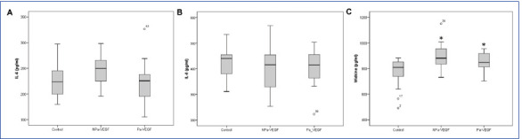
Results: The study included a total of 49 eyes from 49 patients undergoing PPV. The concentrations of IL-6 and IL-8 in vitreous samples from the NPa-VEGF group (n=15) and the Pa-VEGF group (n=14) were not significantly different compared to the control group (n=20) (p>0.05). However, the vitreous fluid of patients in the NPa-VEGF group exhibited significantly higher MK concentrations compared to the control group (p<0.007). Similarly, MK concentrations were significantly elevated in the Pa-VEGF group compared to the control group (p<0.046). No significant difference in MK levels was detected between the NPa-VEGF and Pa-VEGF groups (p>0.05).
Conclusion: These findings suggest that increased MK expression in the vitreous may be associated with the pathogenesis of PDR. Further studies are warranted to elucidate the precise mechanisms underlying this association and to explore the potential of MK as a therapeutic target for PDR management.

 求助内容:
求助内容: 应助结果提醒方式:


