{"title":"Attenuation Estimation and Acoustic Characterization of Mouse Lymph Node Tumor Using High-frequency Ultrasound.","authors":"Masaaki Omura, Kazuki Maeda, Kazuki Tamura, Kenji Yoshida, Ariunbuyan Sukhbaatar, Tetsuya Kodama, Tadashi Yamaguchi","doi":"10.1007/s11307-025-02007-2","DOIUrl":null,"url":null,"abstract":"<p><strong>Purpose: </strong>Lymph node (LN) biopsy is the gold standard for diagnosing metastasis. While ultrasound imaging is a non-invasive method for real-time LN metastasis diagnosis and tumor assessment, its accuracy depends on operator skill and system settings. Quantitative ultrasound can characterize tissue microstructure changes due to tumors, offering operator-independent parameters, and one of the quantitative ultrasound methods, the backscatter coefficient, is necessary to compensate for tissue attenuation. However, the change in the attenuation coefficient (AC) in the tumor growth is uncertain. Using in vivo high-frequency ultrasound (25 MHz) measurement and scanning acoustic microscopy (80 and 300 MHz) for ex vivo samples, we aim to investigate how tumor growth is linked to the attenuation and acoustic properties such as acoustic impedance and speed of sound related to ultrasonic wave propagation.</p><p><strong>Procedures: </strong>FM3 A-Luc mammary carcinoma cells were inoculated into the subiliac LNs of mice, and tumor progression was monitored over time. Bioluminescence imaging was used to assess tumor growth, while ultrasound measurements focused on estimating AC and other acoustic properties.</p><p><strong>Results: </strong>Results indicated that the mean of AC decreased, and its standard deviation increased as tumors grew, correlating with bioluminescence intensity. Furthermore, acoustic impedance and speed of sound varied between normal and tumor tissues, revealing differences in tissue microstructure from the histopathological images.</p><p><strong>Conclusions: </strong>The finding of a decrease in AC observed with tumor growth may play a crucial role in enhancing the accuracy of quantitative ultrasound on attenuation compensation, potentially improving the differentiation between metastatic and non-metastatic LNs.</p>","PeriodicalId":18760,"journal":{"name":"Molecular Imaging and Biology","volume":" ","pages":"379-388"},"PeriodicalIF":2.5000,"publicationDate":"2025-06-01","publicationTypes":"Journal Article","fieldsOfStudy":null,"isOpenAccess":false,"openAccessPdf":"https://www.ncbi.nlm.nih.gov/pmc/articles/PMC12162808/pdf/","citationCount":"0","resultStr":null,"platform":"Semanticscholar","paperid":null,"PeriodicalName":"Molecular Imaging and Biology","FirstCategoryId":"3","ListUrlMain":"https://doi.org/10.1007/s11307-025-02007-2","RegionNum":4,"RegionCategory":"医学","ArticlePicture":[],"TitleCN":null,"AbstractTextCN":null,"PMCID":null,"EPubDate":"2025/5/12 0:00:00","PubModel":"Epub","JCR":"Q2","JCRName":"RADIOLOGY, NUCLEAR MEDICINE & MEDICAL IMAGING","Score":null,"Total":0}
引用次数: 0
Abstract
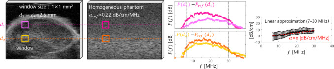
Purpose: Lymph node (LN) biopsy is the gold standard for diagnosing metastasis. While ultrasound imaging is a non-invasive method for real-time LN metastasis diagnosis and tumor assessment, its accuracy depends on operator skill and system settings. Quantitative ultrasound can characterize tissue microstructure changes due to tumors, offering operator-independent parameters, and one of the quantitative ultrasound methods, the backscatter coefficient, is necessary to compensate for tissue attenuation. However, the change in the attenuation coefficient (AC) in the tumor growth is uncertain. Using in vivo high-frequency ultrasound (25 MHz) measurement and scanning acoustic microscopy (80 and 300 MHz) for ex vivo samples, we aim to investigate how tumor growth is linked to the attenuation and acoustic properties such as acoustic impedance and speed of sound related to ultrasonic wave propagation.
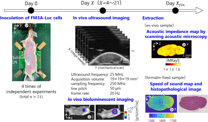
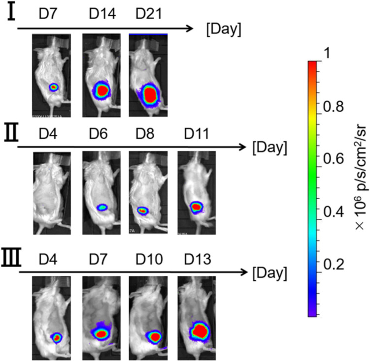
Procedures: FM3 A-Luc mammary carcinoma cells were inoculated into the subiliac LNs of mice, and tumor progression was monitored over time. Bioluminescence imaging was used to assess tumor growth, while ultrasound measurements focused on estimating AC and other acoustic properties.
Results: Results indicated that the mean of AC decreased, and its standard deviation increased as tumors grew, correlating with bioluminescence intensity. Furthermore, acoustic impedance and speed of sound varied between normal and tumor tissues, revealing differences in tissue microstructure from the histopathological images.
Conclusions: The finding of a decrease in AC observed with tumor growth may play a crucial role in enhancing the accuracy of quantitative ultrasound on attenuation compensation, potentially improving the differentiation between metastatic and non-metastatic LNs.
期刊介绍:
Molecular Imaging and Biology (MIB) invites original contributions (research articles, review articles, commentaries, etc.) on the utilization of molecular imaging (i.e., nuclear imaging, optical imaging, autoradiography and pathology, MRI, MPI, ultrasound imaging, radiomics/genomics etc.) to investigate questions related to biology and health. The objective of MIB is to provide a forum to the discovery of molecular mechanisms of disease through the use of imaging techniques. We aim to investigate the biological nature of disease in patients and establish new molecular imaging diagnostic and therapy procedures.
Some areas that are covered are:
Preclinical and clinical imaging of macromolecular targets (e.g., genes, receptors, enzymes) involved in significant biological processes.
The design, characterization, and study of new molecular imaging probes and contrast agents for the functional interrogation of macromolecular targets.
Development and evaluation of imaging systems including instrumentation, image reconstruction algorithms, image analysis, and display.
Development of molecular assay approaches leading to quantification of the biological information obtained in molecular imaging.
Study of in vivo animal models of disease for the development of new molecular diagnostics and therapeutics.
Extension of in vitro and in vivo discoveries using disease models, into well designed clinical research investigations.
Clinical molecular imaging involving clinical investigations, clinical trials and medical management or cost-effectiveness studies.

 求助内容:
求助内容: 应助结果提醒方式:


