Clinson C Lui, Dominique J Wiener, Katia R Groch, Lauren W Stranahan, Christina R Heard, Aníbal G Armién
{"title":"Evaluation of canine epidermis and keratinocytoids (keratinocytic organoids) by transmission electron microscopy.","authors":"Clinson C Lui, Dominique J Wiener, Katia R Groch, Lauren W Stranahan, Christina R Heard, Aníbal G Armién","doi":"10.1111/vde.13356","DOIUrl":null,"url":null,"abstract":"<p><strong>Background: </strong>Skin disease is a common complaint in veterinary medicine. Current models often use live animals. Epidermal organoids (keratinocytoids) are 3D miniature organs created in culture from single epidermal keratinocytes. These keratinocytoids can be used as an alternative to live animal studies to investigate epidermal structures and skin diseases.</p><p><strong>Hypothesis/objective: </strong>This study's objective was to compare ultrastructural morphological features of canine keratinocytoids to those features of normal epidermis.</p><p><strong>Results: </strong>Skin and organoids had morphologically similar components, including tight junctions, desmosomes, lamellar bodies and keratin filaments. These structures were similar in both morphology and distribution. Keratinocytes in the organoids had larger and more distinct keratohyalin granules than epidermal keratinocytes and contained small amounts of glycogen. Keratinocytes from the skin showed no glycogen accumulation.</p><p><strong>Conclusions and clinical relevance: </strong>Canine keratinocytoids are a useful model to study canine epidermal disease from a light microscopy and ultrastructural standpoint.</p>","PeriodicalId":23599,"journal":{"name":"Veterinary dermatology","volume":" ","pages":"696-702"},"PeriodicalIF":1.4000,"publicationDate":"2025-10-01","publicationTypes":"Journal Article","fieldsOfStudy":null,"isOpenAccess":false,"openAccessPdf":"https://www.ncbi.nlm.nih.gov/pmc/articles/PMC12420878/pdf/","citationCount":"0","resultStr":null,"platform":"Semanticscholar","paperid":null,"PeriodicalName":"Veterinary dermatology","FirstCategoryId":"97","ListUrlMain":"https://doi.org/10.1111/vde.13356","RegionNum":3,"RegionCategory":"农林科学","ArticlePicture":[],"TitleCN":null,"AbstractTextCN":null,"PMCID":null,"EPubDate":"2025/5/14 0:00:00","PubModel":"Epub","JCR":"Q3","JCRName":"DERMATOLOGY","Score":null,"Total":0}
引用次数: 0
Abstract
Background: Skin disease is a common complaint in veterinary medicine. Current models often use live animals. Epidermal organoids (keratinocytoids) are 3D miniature organs created in culture from single epidermal keratinocytes. These keratinocytoids can be used as an alternative to live animal studies to investigate epidermal structures and skin diseases.
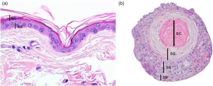
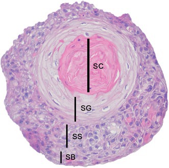
Hypothesis/objective: This study's objective was to compare ultrastructural morphological features of canine keratinocytoids to those features of normal epidermis.
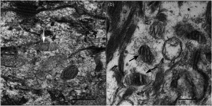
Results: Skin and organoids had morphologically similar components, including tight junctions, desmosomes, lamellar bodies and keratin filaments. These structures were similar in both morphology and distribution. Keratinocytes in the organoids had larger and more distinct keratohyalin granules than epidermal keratinocytes and contained small amounts of glycogen. Keratinocytes from the skin showed no glycogen accumulation.
Conclusions and clinical relevance: Canine keratinocytoids are a useful model to study canine epidermal disease from a light microscopy and ultrastructural standpoint.
期刊介绍:
Veterinary Dermatology is a bi-monthly, peer-reviewed, international journal which publishes papers on all aspects of the skin of mammals, birds, reptiles, amphibians and fish. Scientific research papers, clinical case reports and reviews covering the following aspects of dermatology will be considered for publication:
-Skin structure (anatomy, histology, ultrastructure)
-Skin function (physiology, biochemistry, pharmacology, immunology, genetics)
-Skin microbiology and parasitology
-Dermatopathology
-Pathogenesis, diagnosis and treatment of skin diseases
-New disease entities
 求助内容:
求助内容: 应助结果提醒方式:


