Prakash Singh, Yogita Khandelwal, Vineet Mishra, Bela Jain
{"title":"Intriguing Encounter: Unveiling Squamous Cell Carcinoma Lung with Rare Bilateral Renal and Pituitary Metastasis on [18F]-FDG PET/CT.","authors":"Prakash Singh, Yogita Khandelwal, Vineet Mishra, Bela Jain","doi":"10.4103/ijnm.ijnm_57_24","DOIUrl":null,"url":null,"abstract":"<p><p>Metastases from squamous cell carcinoma of the lung typically occur in the brain, liver, adrenal glands, bone, and regional lymph nodes. It is exceedingly uncommon to encounter multiple rare sites of metastasis from a single primary neoplasm. Herein, we describe a case of a 44-year-old male diagnosed with squamous cell carcinoma lung with pituitary and renal metastasis detected on <sup>18</sup>F-FDG (Fluorodeoxyglucose) PET/CT. <sup>18</sup>F-FDG PET/CT is the standard of care and is an integral part of the clinical staging of patients with lung cancer. According to published literature, the incidence of symptomatic pituitary and renal metastasis from squamous cell carcinoma lung is rare to find with incidences <1% and 5%, respectively. The revelation of rare sites of metastasis originating from primary squamous cell carcinoma lung, as reported in this case on FDG PET/CT, illuminates the exceptional rarity and intricacies in oncology. The exquisite sensitivity of FDG PET/CT enables the identification of occult metastasis in atypical anatomical locations, presenting a distinct advantage over conventional imaging modalities.</p>","PeriodicalId":45830,"journal":{"name":"Indian Journal of Nuclear Medicine","volume":"39 6","pages":"454-456"},"PeriodicalIF":0.5000,"publicationDate":"2024-11-01","publicationTypes":"Journal Article","fieldsOfStudy":null,"isOpenAccess":false,"openAccessPdf":"https://www.ncbi.nlm.nih.gov/pmc/articles/PMC12020972/pdf/","citationCount":"0","resultStr":null,"platform":"Semanticscholar","paperid":null,"PeriodicalName":"Indian Journal of Nuclear Medicine","FirstCategoryId":"1085","ListUrlMain":"https://doi.org/10.4103/ijnm.ijnm_57_24","RegionNum":0,"RegionCategory":null,"ArticlePicture":[],"TitleCN":null,"AbstractTextCN":null,"PMCID":null,"EPubDate":"2025/3/20 0:00:00","PubModel":"Epub","JCR":"Q4","JCRName":"RADIOLOGY, NUCLEAR MEDICINE & MEDICAL IMAGING","Score":null,"Total":0}
引用次数: 0
Abstract
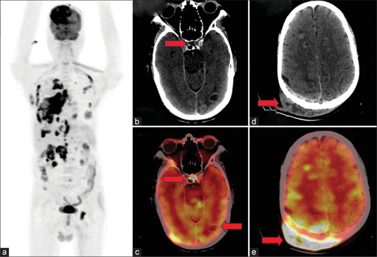
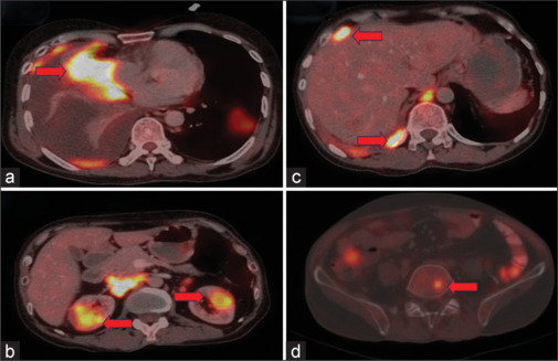
Metastases from squamous cell carcinoma of the lung typically occur in the brain, liver, adrenal glands, bone, and regional lymph nodes. It is exceedingly uncommon to encounter multiple rare sites of metastasis from a single primary neoplasm. Herein, we describe a case of a 44-year-old male diagnosed with squamous cell carcinoma lung with pituitary and renal metastasis detected on 18F-FDG (Fluorodeoxyglucose) PET/CT. 18F-FDG PET/CT is the standard of care and is an integral part of the clinical staging of patients with lung cancer. According to published literature, the incidence of symptomatic pituitary and renal metastasis from squamous cell carcinoma lung is rare to find with incidences <1% and 5%, respectively. The revelation of rare sites of metastasis originating from primary squamous cell carcinoma lung, as reported in this case on FDG PET/CT, illuminates the exceptional rarity and intricacies in oncology. The exquisite sensitivity of FDG PET/CT enables the identification of occult metastasis in atypical anatomical locations, presenting a distinct advantage over conventional imaging modalities.

 求助内容:
求助内容: 应助结果提醒方式:


