Stephan Rau, Thomas Stein, Alexander Rau, Caroline Wilpert, Fabian Pallasch, Balazs Bogner, Sebastian Faby, Fabian Bamberg, Jakob Weiss
{"title":"Simple cystic lesions of the pancreas: image quality and diagnostic accuracy of photon-counting detector computed tomography.","authors":"Stephan Rau, Thomas Stein, Alexander Rau, Caroline Wilpert, Fabian Pallasch, Balazs Bogner, Sebastian Faby, Fabian Bamberg, Jakob Weiss","doi":"10.1007/s11547-025-02015-w","DOIUrl":null,"url":null,"abstract":"<p><strong>Purpose: </strong>To evaluate image quality and diagnostic accuracy of photon-counting detector (PCD) CT for the detection of pancreatic cystic lesions (PCLs) compared to energy-integrating detector (EID) CT with MRI serving as reference standard.</p><p><strong>Material and methods: </strong>We included consecutive patients who underwent contrast-enhanced PCD-CT of the abdomen and for whom an additional abdominal EID-CT was available. Multiparametric MRI served as the reference standard. CT images were assessed for the presence of PCLs by three radiologists independently in a blinded reading. Image quality, lesion conspicuity, and diagnostic confidence were rated on a 5-point Likert scale (5 = excellent). The coefficient of variation (CV) and the density difference between PCLs and visually normal pancreatic parenchyma were calculated as quantitative imaging measures. Radiation dose was assessed using CTDIvol [mGy].</p><p><strong>Results: </strong>Among 106 included patients (age 62.7 ± 12.6 years; 45 [42.5%] male), 46 had MRI-confirmed cystic lesions (mean size 8.7 ± 7.4 mm; range 2-45 mm). Diagnostic accuracy for PCLs was significantly higher for PCD-CT vs. EID-CT (area under the curve: 0.81 vs. 0.74; p = 0.002; sensitivity: 76.8% vs. 59.4%). Image quality, lesion conspicuity, and diagnostic confidence were rated superior for PCD-CT vs. EID-CT (all p < 0.001). Quantitative analyses revealed a significantly lower CV (0.19 vs. 0.24; p = 0.002) and a higher density difference (94.1 HU vs. 76.6 HU p < 0.001) between PCLs and visually normal pancreatic parenchyma at lower radiation doses (7.13 vs. 8.68 mGy; p < 0.001) for PCD-CT vs. EID-CT.</p><p><strong>Conclusion: </strong>PCD-CT provided significantly higher diagnostic accuracy and superior image quality for the detection of PCLs compared to conventional EID-CT at lower radiation dose.</p>","PeriodicalId":20817,"journal":{"name":"Radiologia Medica","volume":" ","pages":"1064-1073"},"PeriodicalIF":4.8000,"publicationDate":"2025-07-01","publicationTypes":"Journal Article","fieldsOfStudy":null,"isOpenAccess":false,"openAccessPdf":"https://www.ncbi.nlm.nih.gov/pmc/articles/PMC12263767/pdf/","citationCount":"0","resultStr":null,"platform":"Semanticscholar","paperid":null,"PeriodicalName":"Radiologia Medica","FirstCategoryId":"3","ListUrlMain":"https://doi.org/10.1007/s11547-025-02015-w","RegionNum":1,"RegionCategory":"医学","ArticlePicture":[],"TitleCN":null,"AbstractTextCN":null,"PMCID":null,"EPubDate":"2025/4/21 0:00:00","PubModel":"Epub","JCR":"Q1","JCRName":"RADIOLOGY, NUCLEAR MEDICINE & MEDICAL IMAGING","Score":null,"Total":0}
引用次数: 0
Abstract
Purpose: To evaluate image quality and diagnostic accuracy of photon-counting detector (PCD) CT for the detection of pancreatic cystic lesions (PCLs) compared to energy-integrating detector (EID) CT with MRI serving as reference standard.
Material and methods: We included consecutive patients who underwent contrast-enhanced PCD-CT of the abdomen and for whom an additional abdominal EID-CT was available. Multiparametric MRI served as the reference standard. CT images were assessed for the presence of PCLs by three radiologists independently in a blinded reading. Image quality, lesion conspicuity, and diagnostic confidence were rated on a 5-point Likert scale (5 = excellent). The coefficient of variation (CV) and the density difference between PCLs and visually normal pancreatic parenchyma were calculated as quantitative imaging measures. Radiation dose was assessed using CTDIvol [mGy].
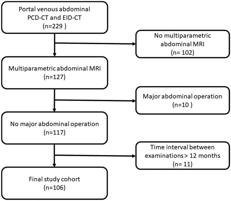
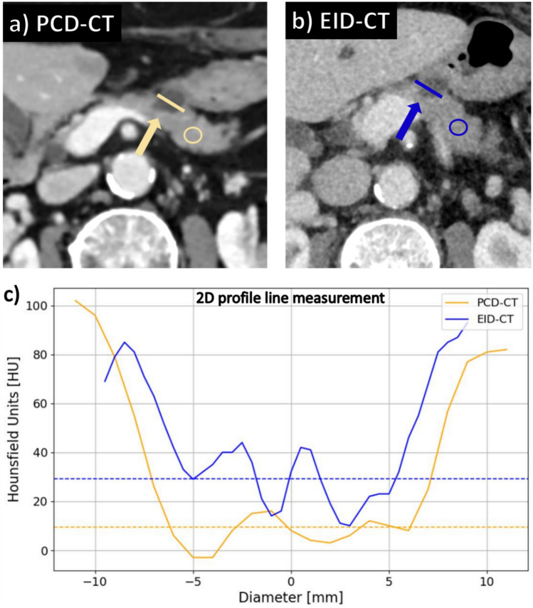
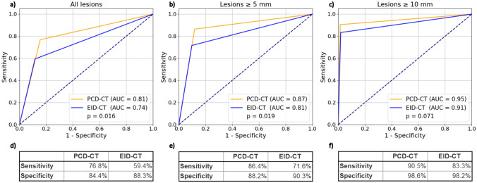
Results: Among 106 included patients (age 62.7 ± 12.6 years; 45 [42.5%] male), 46 had MRI-confirmed cystic lesions (mean size 8.7 ± 7.4 mm; range 2-45 mm). Diagnostic accuracy for PCLs was significantly higher for PCD-CT vs. EID-CT (area under the curve: 0.81 vs. 0.74; p = 0.002; sensitivity: 76.8% vs. 59.4%). Image quality, lesion conspicuity, and diagnostic confidence were rated superior for PCD-CT vs. EID-CT (all p < 0.001). Quantitative analyses revealed a significantly lower CV (0.19 vs. 0.24; p = 0.002) and a higher density difference (94.1 HU vs. 76.6 HU p < 0.001) between PCLs and visually normal pancreatic parenchyma at lower radiation doses (7.13 vs. 8.68 mGy; p < 0.001) for PCD-CT vs. EID-CT.
Conclusion: PCD-CT provided significantly higher diagnostic accuracy and superior image quality for the detection of PCLs compared to conventional EID-CT at lower radiation dose.
期刊介绍:
Felice Perussia founded La radiologia medica in 1914. It is a peer-reviewed journal and serves as the official journal of the Italian Society of Medical and Interventional Radiology (SIRM). The primary purpose of the journal is to disseminate information related to Radiology, especially advancements in diagnostic imaging and related disciplines. La radiologia medica welcomes original research on both fundamental and clinical aspects of modern radiology, with a particular focus on diagnostic and interventional imaging techniques. It also covers topics such as radiotherapy, nuclear medicine, radiobiology, health physics, and artificial intelligence in the context of clinical implications. The journal includes various types of contributions such as original articles, review articles, editorials, short reports, and letters to the editor. With an esteemed Editorial Board and a selection of insightful reports, the journal is an indispensable resource for radiologists and professionals in related fields. Ultimately, La radiologia medica aims to serve as a platform for international collaboration and knowledge sharing within the radiological community.

 求助内容:
求助内容: 应助结果提醒方式:


