Ettore Silvagni, Antonio Marangoni, Carlo Garaffoni, Simone Appenzeller, George Bertsias, Antonis Fanouriakis, Matteo Piga, Enrico Fainardi, Greta Carrara, Carlo Alberto Scirè, Marcello Govoni, Alessandra Bortoluzzi
{"title":"Can conventional brain MRI support the attribution process in neuropsychiatric SLE? A multicentre retrospective study.","authors":"Ettore Silvagni, Antonio Marangoni, Carlo Garaffoni, Simone Appenzeller, George Bertsias, Antonis Fanouriakis, Matteo Piga, Enrico Fainardi, Greta Carrara, Carlo Alberto Scirè, Marcello Govoni, Alessandra Bortoluzzi","doi":"10.1136/lupus-2024-001490","DOIUrl":null,"url":null,"abstract":"<p><strong>Objectives: </strong>We aimed to investigate which elementary lesions, identified through conventional brain MRI, correlated with the attribution of neuropsychiatric (NP) manifestations of SLE as determined by clinical judgement (CJ) and a validated attribution algorithm (AA).</p><p><strong>Methods: </strong>We conducted a multicentre, retrospective cohort study of patients with SLE (1999-2018) from four tertiary SLE centres. Patients were assessed using American College of Rheumatology nomenclature and underwent MRI at their first NP event. NP manifestations were attributed to SLE using CJ and the AA. Elementary lesions were classified as follows: large infarcts, parenchymal haemorrhages, subarachnoid haemorrhages, inflammatory-type lesions, myelopathy, T2/fluid-attenuating inversion recovery (FLAIR) hyperintense lesions, lacunes, cerebral atrophy and microbleeds. Statistical analyses were performed using χ<sup>2</sup> and Fisher's exact tests. Univariable and multivariable logistic regression models were performed. A sensitivity analysis was performed using a revised AA, which excluded the item 'presence of abnormal MRI' from the list of favouring factors.</p><p><strong>Results: </strong>Among 154 patients, 88 (57%) had NP events attributed to SLE by CJ and 85 (55%) by AA. MRI was normal in 57/154 (37%) cases, while T2/FLAIR hyperintense lesions were the most frequent findings (71/154, 46%). A normal MRI was more common in non-attributed NP events per CJ and AA (OR 0.42, 95% CI 0.21 to 0.82 and 0.27, 95% CI 0.13 to 0.52, respectively). Cerebral atrophy was more frequent in non-attributed events per CJ (adjusted OR 0.06, 95% CI 0.01 to 0.35), while inflammatory-type lesions were more prevalent in SLE-attributed events according to AA (OR 3.91, 95% CI 1.15 to 18.1), with no significant change in sensitivity analyses.</p><p><strong>Conclusions: </strong>Our study elucidates the role of conventional MRI findings in the attribution process in NPSLE. The presence of selected elementary lesions or, instead, their absence could have a relevant weight in assessing NP events. These findings may assist clinicians in achieving a more accurate attribution of NP manifestations.</p>","PeriodicalId":18126,"journal":{"name":"Lupus Science & Medicine","volume":"12 1","pages":""},"PeriodicalIF":3.5000,"publicationDate":"2025-04-28","publicationTypes":"Journal Article","fieldsOfStudy":null,"isOpenAccess":false,"openAccessPdf":"https://www.ncbi.nlm.nih.gov/pmc/articles/PMC12039020/pdf/","citationCount":"0","resultStr":null,"platform":"Semanticscholar","paperid":null,"PeriodicalName":"Lupus Science & Medicine","FirstCategoryId":"3","ListUrlMain":"https://doi.org/10.1136/lupus-2024-001490","RegionNum":2,"RegionCategory":"医学","ArticlePicture":[],"TitleCN":null,"AbstractTextCN":null,"PMCID":null,"EPubDate":"","PubModel":"","JCR":"Q1","JCRName":"RHEUMATOLOGY","Score":null,"Total":0}
引用次数: 0
Abstract
Objectives: We aimed to investigate which elementary lesions, identified through conventional brain MRI, correlated with the attribution of neuropsychiatric (NP) manifestations of SLE as determined by clinical judgement (CJ) and a validated attribution algorithm (AA).
Methods: We conducted a multicentre, retrospective cohort study of patients with SLE (1999-2018) from four tertiary SLE centres. Patients were assessed using American College of Rheumatology nomenclature and underwent MRI at their first NP event. NP manifestations were attributed to SLE using CJ and the AA. Elementary lesions were classified as follows: large infarcts, parenchymal haemorrhages, subarachnoid haemorrhages, inflammatory-type lesions, myelopathy, T2/fluid-attenuating inversion recovery (FLAIR) hyperintense lesions, lacunes, cerebral atrophy and microbleeds. Statistical analyses were performed using χ2 and Fisher's exact tests. Univariable and multivariable logistic regression models were performed. A sensitivity analysis was performed using a revised AA, which excluded the item 'presence of abnormal MRI' from the list of favouring factors.
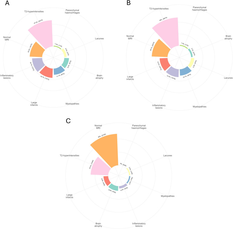
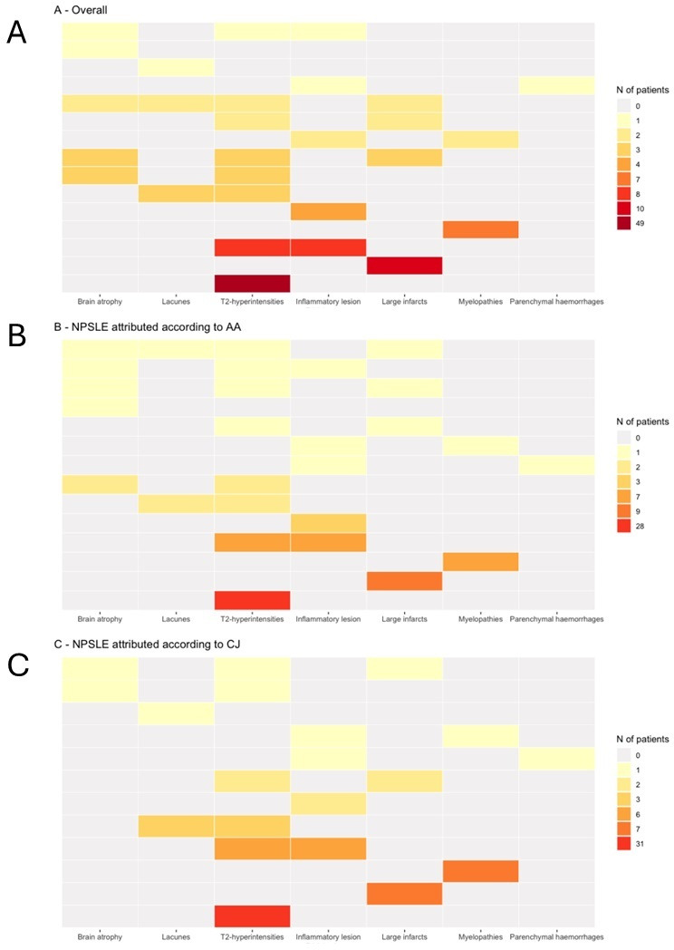
Results: Among 154 patients, 88 (57%) had NP events attributed to SLE by CJ and 85 (55%) by AA. MRI was normal in 57/154 (37%) cases, while T2/FLAIR hyperintense lesions were the most frequent findings (71/154, 46%). A normal MRI was more common in non-attributed NP events per CJ and AA (OR 0.42, 95% CI 0.21 to 0.82 and 0.27, 95% CI 0.13 to 0.52, respectively). Cerebral atrophy was more frequent in non-attributed events per CJ (adjusted OR 0.06, 95% CI 0.01 to 0.35), while inflammatory-type lesions were more prevalent in SLE-attributed events according to AA (OR 3.91, 95% CI 1.15 to 18.1), with no significant change in sensitivity analyses.
Conclusions: Our study elucidates the role of conventional MRI findings in the attribution process in NPSLE. The presence of selected elementary lesions or, instead, their absence could have a relevant weight in assessing NP events. These findings may assist clinicians in achieving a more accurate attribution of NP manifestations.
目的:我们旨在研究通过常规脑MRI识别的哪些初级病变与临床判断(CJ)和经过验证的归因算法(AA)确定的SLE神经精神(NP)表现归因相关。方法:我们对来自4个三级SLE中心的SLE患者(1999-2018)进行了一项多中心、回顾性队列研究。使用美国风湿病学会命名法对患者进行评估,并在首次NP事件时进行MRI检查。使用CJ和AA将NP表现归因于SLE。基础病变分类如下:大梗死、实质出血、蛛网膜下腔出血、炎症型病变、脊髓病、T2/液体衰减反转恢复(FLAIR)高强度病变、凹窝、脑萎缩和微出血。采用χ2和Fisher精确检验进行统计学分析。采用单变量和多变量logistic回归模型。使用修订的AA进行敏感性分析,从有利因素列表中排除了“MRI异常存在”这一项。结果:154例患者中,88例(57%)发生由CJ引起的SLE NP事件,85例(55%)由AA引起。57/154例(37%)MRI正常,而T2/FLAIR高信号病变是最常见的表现(71/154,46%)。MRI正常更常见于每CJ和AA的非归因NP事件(分别为0.42,95% CI 0.21 ~ 0.82和0.27,95% CI 0.13 ~ 0.52)。脑萎缩在每名患者的非归因事件中更为常见(校正OR 0.06, 95% CI 0.01至0.35),而炎症型病变在AA组的sled归因事件中更为普遍(OR 3.91, 95% CI 1.15至18.1),敏感性分析无显著变化。结论:本研究阐明了常规MRI结果在NPSLE归因过程中的作用。所选择的初级病变的存在或不存在可能在评估NP事件中具有相关权重。这些发现可能有助于临床医生获得更准确的NP表现归因。
期刊介绍:
Lupus Science & Medicine is a global, peer reviewed, open access online journal that provides a central point for publication of basic, clinical, translational, and epidemiological studies of all aspects of lupus and related diseases. It is the first lupus-specific open access journal in the world and was developed in response to the need for a barrier-free forum for publication of groundbreaking studies in lupus. The journal publishes research on lupus from fields including, but not limited to: rheumatology, dermatology, nephrology, immunology, pediatrics, cardiology, hepatology, pulmonology, obstetrics and gynecology, and psychiatry.
 求助内容:
求助内容: 应助结果提醒方式:


