Shashi B Singh, Om H Gandhi, Bimash B Shrestha, Patrick Glennan, Anuradha Rosario Bahadur, Niloofaralsadat Motamedi, Kishor Khanal, Sagar Wagle, Poul Flemming Høilund-Carlsen, Thomas J Werner, Mona-Elisabeth Revheim, Abass Alavi
{"title":"[<sup>18</sup>F]NaF PET/CT Imaging of Iliac Bones to Assess Bone Turnover.","authors":"Shashi B Singh, Om H Gandhi, Bimash B Shrestha, Patrick Glennan, Anuradha Rosario Bahadur, Niloofaralsadat Motamedi, Kishor Khanal, Sagar Wagle, Poul Flemming Høilund-Carlsen, Thomas J Werner, Mona-Elisabeth Revheim, Abass Alavi","doi":"10.1007/s11307-025-02003-6","DOIUrl":null,"url":null,"abstract":"<p><strong>Purpose: </strong>This study investigated the effects of laterality, age, gender, BMI, and physical activity level on iliac bone turnover using [<sup>18</sup>F]NaF PET/CT.</p><p><strong>Procedures: </strong>Fifty-nine males and 44 females from the CAMONA study were analyzed. A region of interest (ROI) was drawn to segment the iliac bone using Hounsfield unit thresholds and morphological closing algorithm. [<sup>18</sup>F]NaF SUVmean was compared between the left and right iliac bones using a paired t-test, while Pearson correlation coefficient assessed changes with age, BMI, and physical activity level.</p><p><strong>Results: </strong>[<sup>18</sup>F]NaF uptake was higher in right iliac bone than left in males, females, and the combined-group. In males, SUVmean was 2.98 ± 1.63 (1.1-7.87) on left and 3.71 ± 1.49 (1.49-3.7) on right. In females, SUVmean was 2.59 ± 1.14 (0.88-6.27) on left and 3.72 ± 1.04 (2.22-6.51) on right. Combined, SUVmean was 2.81 ± 1.44 (0.88-7.87) on left and 3.71 ± 1.31 (0.89-8.07) on right. [<sup>18</sup>F]NaF uptake negatively correlated with age (right: r = - 0.27, P = 0.006; left: r = - 0.22, P = 0.02), stronger in females (right: r = - 0.30, P = 0.04; left: r = - 0.31, P = 0.04) than males (right: r = - 0.26, P = 0.04; left: r = - 0.18, P = 0.18). SUVmean correlated positively with BMI in males (right: r = 0.47, P = 0.0002; left: r = 0.38, P = 0.0027), females (right: r = 0.36, P = 0.0168; left: r = 0.30, P = 0.0505), and combined-group (right: r = 0.43, P < 0.0001; left: r = 0.37, P = 0.0001). No significant correlation was found between SUVmean and physical activity in males, while in females, a negative correlation was observed on left (r = - 0.37, P = 0.0390) but not on right (r = - 0.27, P = 0.1302), and when combined, the correlation remained significant on left (r = - 0.24, P = 0.0372) but not on right (r = - 0.16, P = 0.1541).</p><p><strong>Conclusions: </strong>[<sup>18</sup>F]NaF uptake was higher in the right iliac bone and declined with age, particularly in females. The positive correlation between SUVmean and BMI; and the negative correlation between SUVmean and physical activity suggest metabolic influences on bone turnover. [<sup>18</sup>F]NaF PET/CT may serve as a tool for assessing bone metabolism and turnover in asymptomatic individuals.</p>","PeriodicalId":18760,"journal":{"name":"Molecular Imaging and Biology","volume":" ","pages":"295-304"},"PeriodicalIF":2.5000,"publicationDate":"2025-06-01","publicationTypes":"Journal Article","fieldsOfStudy":null,"isOpenAccess":false,"openAccessPdf":"https://www.ncbi.nlm.nih.gov/pmc/articles/PMC12162772/pdf/","citationCount":"0","resultStr":null,"platform":"Semanticscholar","paperid":null,"PeriodicalName":"Molecular Imaging and Biology","FirstCategoryId":"3","ListUrlMain":"https://doi.org/10.1007/s11307-025-02003-6","RegionNum":4,"RegionCategory":"医学","ArticlePicture":[],"TitleCN":null,"AbstractTextCN":null,"PMCID":null,"EPubDate":"2025/4/24 0:00:00","PubModel":"Epub","JCR":"Q2","JCRName":"RADIOLOGY, NUCLEAR MEDICINE & MEDICAL IMAGING","Score":null,"Total":0}
引用次数: 0
Abstract
Purpose: This study investigated the effects of laterality, age, gender, BMI, and physical activity level on iliac bone turnover using [18F]NaF PET/CT.
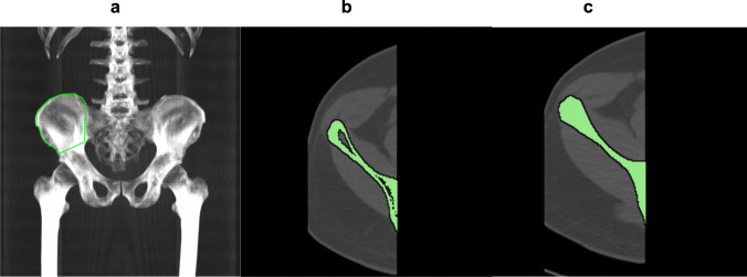
Procedures: Fifty-nine males and 44 females from the CAMONA study were analyzed. A region of interest (ROI) was drawn to segment the iliac bone using Hounsfield unit thresholds and morphological closing algorithm. [18F]NaF SUVmean was compared between the left and right iliac bones using a paired t-test, while Pearson correlation coefficient assessed changes with age, BMI, and physical activity level.
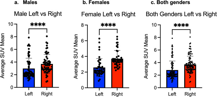
Results: [18F]NaF uptake was higher in right iliac bone than left in males, females, and the combined-group. In males, SUVmean was 2.98 ± 1.63 (1.1-7.87) on left and 3.71 ± 1.49 (1.49-3.7) on right. In females, SUVmean was 2.59 ± 1.14 (0.88-6.27) on left and 3.72 ± 1.04 (2.22-6.51) on right. Combined, SUVmean was 2.81 ± 1.44 (0.88-7.87) on left and 3.71 ± 1.31 (0.89-8.07) on right. [18F]NaF uptake negatively correlated with age (right: r = - 0.27, P = 0.006; left: r = - 0.22, P = 0.02), stronger in females (right: r = - 0.30, P = 0.04; left: r = - 0.31, P = 0.04) than males (right: r = - 0.26, P = 0.04; left: r = - 0.18, P = 0.18). SUVmean correlated positively with BMI in males (right: r = 0.47, P = 0.0002; left: r = 0.38, P = 0.0027), females (right: r = 0.36, P = 0.0168; left: r = 0.30, P = 0.0505), and combined-group (right: r = 0.43, P < 0.0001; left: r = 0.37, P = 0.0001). No significant correlation was found between SUVmean and physical activity in males, while in females, a negative correlation was observed on left (r = - 0.37, P = 0.0390) but not on right (r = - 0.27, P = 0.1302), and when combined, the correlation remained significant on left (r = - 0.24, P = 0.0372) but not on right (r = - 0.16, P = 0.1541).
Conclusions: [18F]NaF uptake was higher in the right iliac bone and declined with age, particularly in females. The positive correlation between SUVmean and BMI; and the negative correlation between SUVmean and physical activity suggest metabolic influences on bone turnover. [18F]NaF PET/CT may serve as a tool for assessing bone metabolism and turnover in asymptomatic individuals.
期刊介绍:
Molecular Imaging and Biology (MIB) invites original contributions (research articles, review articles, commentaries, etc.) on the utilization of molecular imaging (i.e., nuclear imaging, optical imaging, autoradiography and pathology, MRI, MPI, ultrasound imaging, radiomics/genomics etc.) to investigate questions related to biology and health. The objective of MIB is to provide a forum to the discovery of molecular mechanisms of disease through the use of imaging techniques. We aim to investigate the biological nature of disease in patients and establish new molecular imaging diagnostic and therapy procedures.
Some areas that are covered are:
Preclinical and clinical imaging of macromolecular targets (e.g., genes, receptors, enzymes) involved in significant biological processes.
The design, characterization, and study of new molecular imaging probes and contrast agents for the functional interrogation of macromolecular targets.
Development and evaluation of imaging systems including instrumentation, image reconstruction algorithms, image analysis, and display.
Development of molecular assay approaches leading to quantification of the biological information obtained in molecular imaging.
Study of in vivo animal models of disease for the development of new molecular diagnostics and therapeutics.
Extension of in vitro and in vivo discoveries using disease models, into well designed clinical research investigations.
Clinical molecular imaging involving clinical investigations, clinical trials and medical management or cost-effectiveness studies.


 求助内容:
求助内容: 应助结果提醒方式:


