A. A. Sheader, G Vizcay-Barrena, R. A. Fleck, S. J. L. Flatters, P. D. Nellist
{"title":"Subcellular localisation and identification of single atoms using quantitative scanning transmission electron microscopy","authors":"A. A. Sheader, G Vizcay-Barrena, R. A. Fleck, S. J. L. Flatters, P. D. Nellist","doi":"10.1111/jmi.13410","DOIUrl":null,"url":null,"abstract":"<p>Determining the concentration of elements in subcellular structures poses a significant challenge. By locating an elemental species at high spatial resolution and with subcellular context, and subsequently quantifying it on an absolute scale, new information about cellular function can be revealed. Such measurements have not as yet been realised with existing techniques due to limitations on spatial resolution and inherent difficulties in detecting elements present in low concentrations. In this paper, we use scanning transmission electron microscopy (STEM) to establish a methodology for localising and quantifying high-<i>Z</i> elements in a biological setting by measuring elastic electron scattering. We demonstrate platinum (Pt) deposition within neuronal cell bodies following in vivo administration of the Pt-based chemotherapeutic oxaliplatin to validate this novel methodology. For the first time, individual Pt atoms and nanoscale Pt clusters are shown within subcellular structures. Quantitative measurements of elastic electron scattering are used to determine absolute numbers of Pt atoms in each cluster. Cluster density is calculated on an atoms-per-cubic-nanometre scale, and used to show clusters form with densities below that of metallic Pt. By considering STEM partial scattering cross-sections, we determine that this new approach to subcellular elemental detection may be applicable to elements as light as sodium.</p><p><b>LAY DESCRIPTION</b>: Heterogeneous elemental distributions drive fundamental biological processes within cells. While carbon, hydrogen, oxygen and nitrogen comprise by far the majority of living matter, concentrations and locations of more than a dozen other species must also be tightly controlled to ensure normal cell function. Oxaliplatin is a first-line and adjuvant treatment for colorectal cancer. However, pain in the body's extremities (fingers and toes) significantly impairs clinical usage as this serious and persistent side effect impacts on both patient cancer care and quality of life. Annular dark-field (ADF) imaging in the scanning transmission electron microscope (STEM) provides an image with strong atom-number contrast and is sufficient to distinguish between different cell types and different organelles within the cells of the DRG. We also show that Pt may be imaged at the single atom level and be localised at very high resolution while still preserving a degree of ultrastructural context. The intrinsic image contrast generated is sufficient to identify these features without the need for heavy metal stains and other extensive processing steps which risk disturbing native platinum distributions within the tissue. We subsequently demonstrate that by considering the total elastic scattering intensity generated by nanometre-sized Pt aggregations within the cell, the ADF STEM may be used to make a measurement of local concentration of Pt in units of atoms per cubic nanometre. We further estimate the minimum atomic number required to visualise single atoms in this setting, concluding that in similar samples it may be possible to detect species as light as sodium with atomic sensitivity.</p>","PeriodicalId":16484,"journal":{"name":"Journal of microscopy","volume":"299 1","pages":"36-48"},"PeriodicalIF":1.9000,"publicationDate":"2025-04-15","publicationTypes":"Journal Article","fieldsOfStudy":null,"isOpenAccess":false,"openAccessPdf":"https://onlinelibrary.wiley.com/doi/epdf/10.1111/jmi.13410","citationCount":"0","resultStr":null,"platform":"Semanticscholar","paperid":null,"PeriodicalName":"Journal of microscopy","FirstCategoryId":"5","ListUrlMain":"https://onlinelibrary.wiley.com/doi/10.1111/jmi.13410","RegionNum":4,"RegionCategory":"工程技术","ArticlePicture":[],"TitleCN":null,"AbstractTextCN":null,"PMCID":null,"EPubDate":"","PubModel":"","JCR":"Q3","JCRName":"MICROSCOPY","Score":null,"Total":0}
引用次数: 0
Abstract
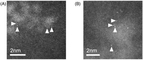
Determining the concentration of elements in subcellular structures poses a significant challenge. By locating an elemental species at high spatial resolution and with subcellular context, and subsequently quantifying it on an absolute scale, new information about cellular function can be revealed. Such measurements have not as yet been realised with existing techniques due to limitations on spatial resolution and inherent difficulties in detecting elements present in low concentrations. In this paper, we use scanning transmission electron microscopy (STEM) to establish a methodology for localising and quantifying high-Z elements in a biological setting by measuring elastic electron scattering. We demonstrate platinum (Pt) deposition within neuronal cell bodies following in vivo administration of the Pt-based chemotherapeutic oxaliplatin to validate this novel methodology. For the first time, individual Pt atoms and nanoscale Pt clusters are shown within subcellular structures. Quantitative measurements of elastic electron scattering are used to determine absolute numbers of Pt atoms in each cluster. Cluster density is calculated on an atoms-per-cubic-nanometre scale, and used to show clusters form with densities below that of metallic Pt. By considering STEM partial scattering cross-sections, we determine that this new approach to subcellular elemental detection may be applicable to elements as light as sodium.
LAY DESCRIPTION: Heterogeneous elemental distributions drive fundamental biological processes within cells. While carbon, hydrogen, oxygen and nitrogen comprise by far the majority of living matter, concentrations and locations of more than a dozen other species must also be tightly controlled to ensure normal cell function. Oxaliplatin is a first-line and adjuvant treatment for colorectal cancer. However, pain in the body's extremities (fingers and toes) significantly impairs clinical usage as this serious and persistent side effect impacts on both patient cancer care and quality of life. Annular dark-field (ADF) imaging in the scanning transmission electron microscope (STEM) provides an image with strong atom-number contrast and is sufficient to distinguish between different cell types and different organelles within the cells of the DRG. We also show that Pt may be imaged at the single atom level and be localised at very high resolution while still preserving a degree of ultrastructural context. The intrinsic image contrast generated is sufficient to identify these features without the need for heavy metal stains and other extensive processing steps which risk disturbing native platinum distributions within the tissue. We subsequently demonstrate that by considering the total elastic scattering intensity generated by nanometre-sized Pt aggregations within the cell, the ADF STEM may be used to make a measurement of local concentration of Pt in units of atoms per cubic nanometre. We further estimate the minimum atomic number required to visualise single atoms in this setting, concluding that in similar samples it may be possible to detect species as light as sodium with atomic sensitivity.
期刊介绍:
The Journal of Microscopy is the oldest journal dedicated to the science of microscopy and the only peer-reviewed publication of the Royal Microscopical Society. It publishes papers that report on the very latest developments in microscopy such as advances in microscopy techniques or novel areas of application. The Journal does not seek to publish routine applications of microscopy or specimen preparation even though the submission may otherwise have a high scientific merit.
The scope covers research in the physical and biological sciences and covers imaging methods using light, electrons, X-rays and other radiations as well as atomic force and near field techniques. Interdisciplinary research is welcome. Papers pertaining to microscopy are also welcomed on optical theory, spectroscopy, novel specimen preparation and manipulation methods and image recording, processing and analysis including dynamic analysis of living specimens.
Publication types include full papers, hot topic fast tracked communications and review articles. Authors considering submitting a review article should contact the editorial office first.

 求助内容:
求助内容: 应助结果提醒方式:


