Suneela Shaukat, Ali Mansoor, Nawaz Rashid, Zara Shaukat, Umar Amin, Sobia Mazhar
{"title":"Diagnostic accuracy of diffusion-weighted magnetic resonance imaging for cervical lymph node metastasis from oral cancer.","authors":"Suneela Shaukat, Ali Mansoor, Nawaz Rashid, Zara Shaukat, Umar Amin, Sobia Mazhar","doi":"10.1590/0100-3984.2024.0064","DOIUrl":null,"url":null,"abstract":"<p><strong>Objective: </strong>To determine the accuracy of diffusion-weighted imaging (DWI) for the diagnosis of cervical lymph node metastasis from oral cancer.</p><p><strong>Materials and methods: </strong>This was a cross-sectional study conducted in the Radiology Department of the Mayo Hospital, in the city of Lahore, Pakistan. We included 150 patients diagnosed with oral cancer. Ages ranged from 18 to 60 years of age. During the study period, all of the patients included underwent magnetic resonance imaging, including a DWI sequence, in a 1.5-T scanner with a phased-array head and neck coil. Patients with contraindications to magnetic resonance (aneurysm, a pacemaker, clips, plates, a prosthetic valve, or claustrophobia) were excluded. In the DWI sequence, the area scanned included the lymph nodes from suprasternal notch to the base of the skull. Histopathology of the lymph nodes was employed as the gold standard.</p><p><strong>Results: </strong>The sensitivity, specificity, positive predictive value, negative predictive value, and accuracy of DWI for the diagnosis of oral cancer metastasis to cervical lymph nodes, with histopathology as the gold standard, was 90.57%, 91.75%, 94.68%, 90.57%, and 91.33%, respectively.</p><p><strong>Conclusion: </strong>Our findings indicate that DWI is fairly accurate for detecting metastases in the cervical lymph nodes of patients with oral cancer.</p>","PeriodicalId":20842,"journal":{"name":"Radiologia Brasileira","volume":"58 ","pages":"e20240064"},"PeriodicalIF":0.0000,"publicationDate":"2025-04-11","publicationTypes":"Journal Article","fieldsOfStudy":null,"isOpenAccess":false,"openAccessPdf":"https://www.ncbi.nlm.nih.gov/pmc/articles/PMC12005712/pdf/","citationCount":"0","resultStr":null,"platform":"Semanticscholar","paperid":null,"PeriodicalName":"Radiologia Brasileira","FirstCategoryId":"1085","ListUrlMain":"https://doi.org/10.1590/0100-3984.2024.0064","RegionNum":0,"RegionCategory":null,"ArticlePicture":[],"TitleCN":null,"AbstractTextCN":null,"PMCID":null,"EPubDate":"2025/1/1 0:00:00","PubModel":"eCollection","JCR":"Q3","JCRName":"Medicine","Score":null,"Total":0}
引用次数: 0
Abstract
Objective: To determine the accuracy of diffusion-weighted imaging (DWI) for the diagnosis of cervical lymph node metastasis from oral cancer.
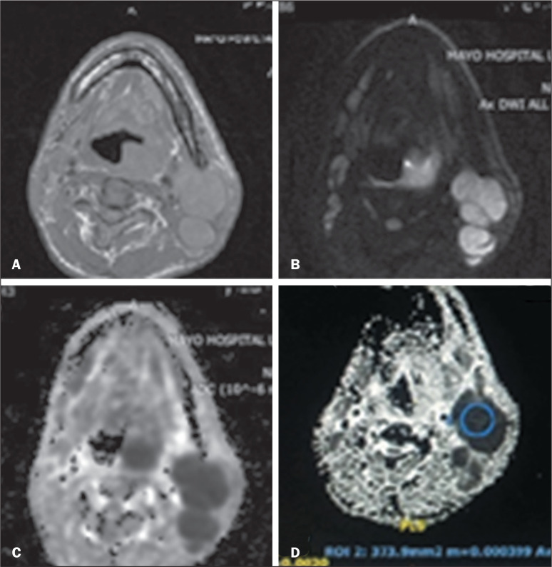
Materials and methods: This was a cross-sectional study conducted in the Radiology Department of the Mayo Hospital, in the city of Lahore, Pakistan. We included 150 patients diagnosed with oral cancer. Ages ranged from 18 to 60 years of age. During the study period, all of the patients included underwent magnetic resonance imaging, including a DWI sequence, in a 1.5-T scanner with a phased-array head and neck coil. Patients with contraindications to magnetic resonance (aneurysm, a pacemaker, clips, plates, a prosthetic valve, or claustrophobia) were excluded. In the DWI sequence, the area scanned included the lymph nodes from suprasternal notch to the base of the skull. Histopathology of the lymph nodes was employed as the gold standard.
Results: The sensitivity, specificity, positive predictive value, negative predictive value, and accuracy of DWI for the diagnosis of oral cancer metastasis to cervical lymph nodes, with histopathology as the gold standard, was 90.57%, 91.75%, 94.68%, 90.57%, and 91.33%, respectively.
Conclusion: Our findings indicate that DWI is fairly accurate for detecting metastases in the cervical lymph nodes of patients with oral cancer.

 求助内容:
求助内容: 应助结果提醒方式:


