Esmée M Bosman, Max E Keizer, Jasper van Aalst, Martinus P G Broen, Alida A Postma, Astrid I P Vernemmen, Henricus P M Kunst, Yasin Temel
{"title":"Spontaneous Shrinking and Growing Skull Base Chordoma.","authors":"Esmée M Bosman, Max E Keizer, Jasper van Aalst, Martinus P G Broen, Alida A Postma, Astrid I P Vernemmen, Henricus P M Kunst, Yasin Temel","doi":"10.1055/a-2587-6573","DOIUrl":null,"url":null,"abstract":"<p><strong>Background: </strong>Chordomas are rare slow-growing tumors occurring in the axial skeleton and can demonstrate local aggressive behavior, typically extending from the median axis, compressing surrounding tissue. Complete surgical resection and adjuvant radiotherapy are the preferred treatments. We present an unusual case of a spontaneously shrinking and growing off-midline petroclival chordoma.</p><p><strong>Case description: </strong>A 23-year-old woman presented with right abducens nerve palsy. Computed tomography and magnetic resonance imaging (MRI) revealed an off-midline petroclival lesion compressing the abducens nerve with characteristics of a chondrosarcoma. Preoperative MRI indicated spontaneous lesion regression, and the abducens nerve showed clinical improvement. Hence, the planned surgery was canceled. During the wait-and-scan period, abducens nerve palsy recurred. MRI confirmed lesion growth and showed an intratumoral linear structure indicative of blood. Even though preoperative MRI again demonstrated shrinkage, the lesion was surgically removed. Despite the unusual presentation, histopathological examination diagnosed a conventional chordoma. A second surgery was required to remove the residual tumor, after which the patient received high-dose photon beam therapy.</p><p><strong>Conclusion: </strong>This article discusses the uncommon presentation and behavior of a petroclival chordoma, showing fluctuating cycles of off-midline growth and spontaneous regression. While intratumoral hemorrhage is hypothesized to explain this tumor behavior, the exact etiology needs further investigation. The case presented here emphasizes the importance of considering chordoma in the differential diagnosis despite an atypical disease course.</p>","PeriodicalId":44256,"journal":{"name":"Journal of Neurological Surgery Reports","volume":"86 2","pages":"e107-e111"},"PeriodicalIF":0.7000,"publicationDate":"2025-05-09","publicationTypes":"Journal Article","fieldsOfStudy":null,"isOpenAccess":false,"openAccessPdf":"https://www.ncbi.nlm.nih.gov/pmc/articles/PMC12064314/pdf/","citationCount":"0","resultStr":null,"platform":"Semanticscholar","paperid":null,"PeriodicalName":"Journal of Neurological Surgery Reports","FirstCategoryId":"1085","ListUrlMain":"https://doi.org/10.1055/a-2587-6573","RegionNum":0,"RegionCategory":null,"ArticlePicture":[],"TitleCN":null,"AbstractTextCN":null,"PMCID":null,"EPubDate":"2025/4/1 0:00:00","PubModel":"eCollection","JCR":"Q4","JCRName":"CLINICAL NEUROLOGY","Score":null,"Total":0}
引用次数: 0
Abstract
Background: Chordomas are rare slow-growing tumors occurring in the axial skeleton and can demonstrate local aggressive behavior, typically extending from the median axis, compressing surrounding tissue. Complete surgical resection and adjuvant radiotherapy are the preferred treatments. We present an unusual case of a spontaneously shrinking and growing off-midline petroclival chordoma.
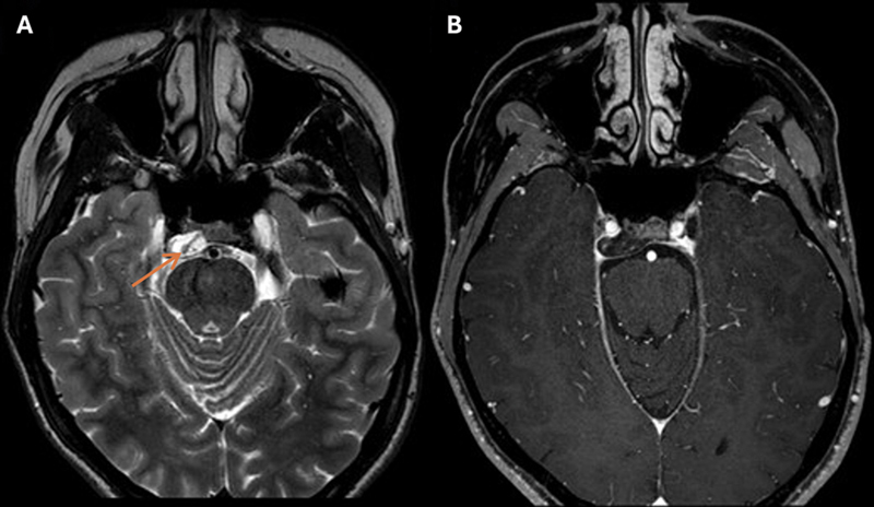
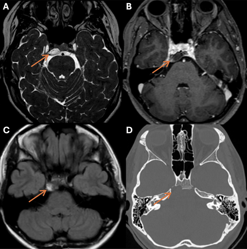
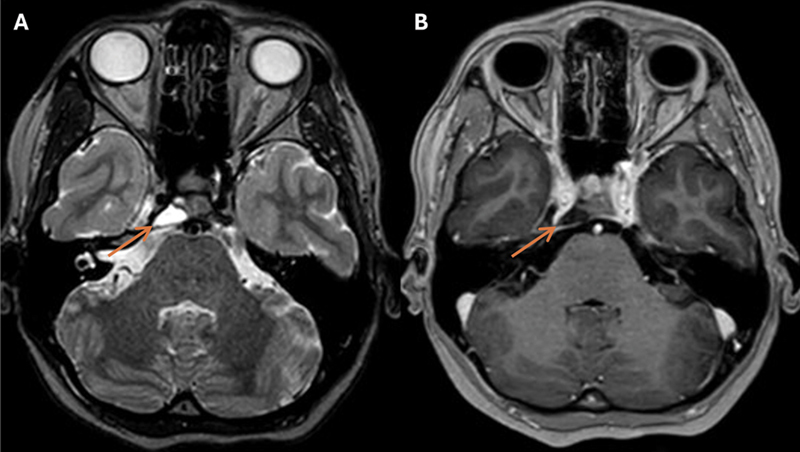
Case description: A 23-year-old woman presented with right abducens nerve palsy. Computed tomography and magnetic resonance imaging (MRI) revealed an off-midline petroclival lesion compressing the abducens nerve with characteristics of a chondrosarcoma. Preoperative MRI indicated spontaneous lesion regression, and the abducens nerve showed clinical improvement. Hence, the planned surgery was canceled. During the wait-and-scan period, abducens nerve palsy recurred. MRI confirmed lesion growth and showed an intratumoral linear structure indicative of blood. Even though preoperative MRI again demonstrated shrinkage, the lesion was surgically removed. Despite the unusual presentation, histopathological examination diagnosed a conventional chordoma. A second surgery was required to remove the residual tumor, after which the patient received high-dose photon beam therapy.
Conclusion: This article discusses the uncommon presentation and behavior of a petroclival chordoma, showing fluctuating cycles of off-midline growth and spontaneous regression. While intratumoral hemorrhage is hypothesized to explain this tumor behavior, the exact etiology needs further investigation. The case presented here emphasizes the importance of considering chordoma in the differential diagnosis despite an atypical disease course.

 求助内容:
求助内容: 应助结果提醒方式:


