Kento Furihata, Jun Miyagawa, Naomichi Tamaru, Hiroyuki Kubota
{"title":"Usefulness of high tube voltage conditions in CT Fluoroscopy during CT-guided biopsy: Preliminary study.","authors":"Kento Furihata, Jun Miyagawa, Naomichi Tamaru, Hiroyuki Kubota","doi":"10.25259/JCIS_7_2025","DOIUrl":null,"url":null,"abstract":"<p><strong>Objectives: </strong>Computed tomography (CT)-guided biopsy is often used to increase the safety and accuracy of biopsies for disease diagnosis. However, CT-guided biopsy is associated with metal artifacts from the biopsy needle and increased patient and operator exposure due to frequent CT fluoroscopy. Therefore, we thought it possible to solve this problem by setting the CT fluoroscopy conditions to a higher tube voltage and a lower tube current-time product (high-tube voltage conditions) than recommended. As a preliminary study, metal artifacts, low-contrast detectability, patient and operator's exposure, and visual changes in high-tube voltage conditions were assessed using phantoms and compared with recommended conditions.</p><p><strong>Material and methods: </strong>On an interventional radiology CT system, the phantom was scanned under recommended conditions (120 kV, 30 mAs) and high-tube voltage conditions (135 kV, 30-5 mAs). The metal artifacts and low-contrast detectability of each condition were analyzed and compared using the acquired images. In addition, the phantom surface dose assuming patient exposure and the air dose assuming the operator's standing position were measured and compared. Furthermore, visual assessment was performed by six radiological technologists.</p><p><strong>Results: </strong>Low-contrast detectability was slightly reduced, metal artifacts were significantly lower under high-tube voltage conditions, and patient and operator exposure were lower than the recommended conditions. Furthermore, the findings of the visual assessment were largely consistent with those of the physical assessment.</p><p><strong>Conclusion: </strong>High-tube voltage conditions in CT fluoroscopy during CT-guided biopsy may be useful in reducing metallic artifacts and patient and operator radiation exposure.</p>","PeriodicalId":15512,"journal":{"name":"Journal of Clinical Imaging Science","volume":"15 ","pages":"13"},"PeriodicalIF":1.3000,"publicationDate":"2025-04-21","publicationTypes":"Journal Article","fieldsOfStudy":null,"isOpenAccess":false,"openAccessPdf":"https://www.ncbi.nlm.nih.gov/pmc/articles/PMC12057212/pdf/","citationCount":"0","resultStr":null,"platform":"Semanticscholar","paperid":null,"PeriodicalName":"Journal of Clinical Imaging Science","FirstCategoryId":"1085","ListUrlMain":"https://doi.org/10.25259/JCIS_7_2025","RegionNum":0,"RegionCategory":null,"ArticlePicture":[],"TitleCN":null,"AbstractTextCN":null,"PMCID":null,"EPubDate":"2025/1/1 0:00:00","PubModel":"eCollection","JCR":"Q3","JCRName":"RADIOLOGY, NUCLEAR MEDICINE & MEDICAL IMAGING","Score":null,"Total":0}
引用次数: 0
Abstract
Objectives: Computed tomography (CT)-guided biopsy is often used to increase the safety and accuracy of biopsies for disease diagnosis. However, CT-guided biopsy is associated with metal artifacts from the biopsy needle and increased patient and operator exposure due to frequent CT fluoroscopy. Therefore, we thought it possible to solve this problem by setting the CT fluoroscopy conditions to a higher tube voltage and a lower tube current-time product (high-tube voltage conditions) than recommended. As a preliminary study, metal artifacts, low-contrast detectability, patient and operator's exposure, and visual changes in high-tube voltage conditions were assessed using phantoms and compared with recommended conditions.
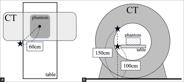
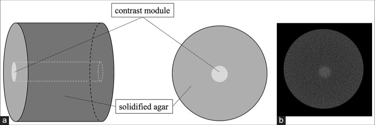
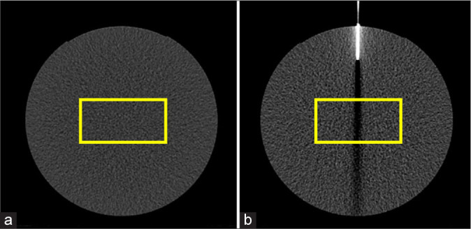
Material and methods: On an interventional radiology CT system, the phantom was scanned under recommended conditions (120 kV, 30 mAs) and high-tube voltage conditions (135 kV, 30-5 mAs). The metal artifacts and low-contrast detectability of each condition were analyzed and compared using the acquired images. In addition, the phantom surface dose assuming patient exposure and the air dose assuming the operator's standing position were measured and compared. Furthermore, visual assessment was performed by six radiological technologists.
Results: Low-contrast detectability was slightly reduced, metal artifacts were significantly lower under high-tube voltage conditions, and patient and operator exposure were lower than the recommended conditions. Furthermore, the findings of the visual assessment were largely consistent with those of the physical assessment.
Conclusion: High-tube voltage conditions in CT fluoroscopy during CT-guided biopsy may be useful in reducing metallic artifacts and patient and operator radiation exposure.
期刊介绍:
The Journal of Clinical Imaging Science (JCIS) is an open access peer-reviewed journal committed to publishing high-quality articles in the field of Imaging Science. The journal aims to present Imaging Science and relevant clinical information in an understandable and useful format. The journal is owned and published by the Scientific Scholar. Audience Our audience includes Radiologists, Researchers, Clinicians, medical professionals and students. Review process JCIS has a highly rigorous peer-review process that makes sure that manuscripts are scientifically accurate, relevant, novel and important. Authors disclose all conflicts, affiliations and financial associations such that the published content is not biased.

 求助内容:
求助内容: 应助结果提醒方式:


