Franziska A Lang, Norbert A Lang, Julia Vorloeper, Christian Niederau, Rogerio B Craveiro, Isabel Knaup, Michael Wolf
{"title":"Validation of a digital, partly automated three-dimensional cast analysis for evaluation of orthodontic treatment assessment.","authors":"Franziska A Lang, Norbert A Lang, Julia Vorloeper, Christian Niederau, Rogerio B Craveiro, Isabel Knaup, Michael Wolf","doi":"10.1186/s13005-025-00515-8","DOIUrl":null,"url":null,"abstract":"<p><strong>Background: </strong>Plaster models have been considered the gold standard in traditional orthodontic model analysis. Modern imaging techniques and ever-advancing technologies have expanded the scope of digital diagnostic tools. These innovations allow the use of devices specifically designed for the diagnosis of 3D structures. The aim of this method comparison study was to determine the accuracy and efficiency of digital measurements compared to conventional manual measurements on plaster models.</p><p><strong>Materials and methods: </strong>The present cohort constitutes the evaluation of pretherapeutic situation models of 247 orthodontically treated patients (129 females and 118 males, average age 16.76 +- 9.49 years) with mixed or permanent dentition who were treated at the University Hospital RWTH Aachen between January 2018 and December 2020. Plaster models were digitised using a model scanner, and an experienced examiner performed various measurements on blinded plaster models using a calliper and on digital models using the specially developed 'Tooth width analysis Aachen' patch in the OnyxCeph3TM-3D software. The intra-rater and inter-rater reliability were determined by a second, blinded assessor. Measurements included tooth width, crown height, arch width, arch length and arch circumference, as well as overjet and overbite. Differences between analogue and digital methods were calculated.</p><p><strong>Results: </strong>Differences of up to 0.3 mm were observed between manual and partially automated digital measurements for sagittal, transversal and vertical parameters. Teeth with close proximal contact to adjacent teeth and teeth in jaws with a negative space analysis result showed an increased difference between manual and partially automated digital measurements, although this was not clinically relevant. The time required to perform digital measurements was statistically significantly reduced.</p><p><strong>Conclusions: </strong>Partially automated digital impression analysis offers an accurate, highly efficient and time-saving alternative to traditional manual impression analysis.</p>","PeriodicalId":12994,"journal":{"name":"Head & Face Medicine","volume":"21 1","pages":"36"},"PeriodicalIF":2.4000,"publicationDate":"2025-05-08","publicationTypes":"Journal Article","fieldsOfStudy":null,"isOpenAccess":false,"openAccessPdf":"https://www.ncbi.nlm.nih.gov/pmc/articles/PMC12060358/pdf/","citationCount":"0","resultStr":null,"platform":"Semanticscholar","paperid":null,"PeriodicalName":"Head & Face Medicine","FirstCategoryId":"3","ListUrlMain":"https://doi.org/10.1186/s13005-025-00515-8","RegionNum":2,"RegionCategory":"医学","ArticlePicture":[],"TitleCN":null,"AbstractTextCN":null,"PMCID":null,"EPubDate":"","PubModel":"","JCR":"Q2","JCRName":"DENTISTRY, ORAL SURGERY & MEDICINE","Score":null,"Total":0}
引用次数: 0
Abstract
Background: Plaster models have been considered the gold standard in traditional orthodontic model analysis. Modern imaging techniques and ever-advancing technologies have expanded the scope of digital diagnostic tools. These innovations allow the use of devices specifically designed for the diagnosis of 3D structures. The aim of this method comparison study was to determine the accuracy and efficiency of digital measurements compared to conventional manual measurements on plaster models.
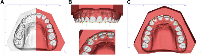
Materials and methods: The present cohort constitutes the evaluation of pretherapeutic situation models of 247 orthodontically treated patients (129 females and 118 males, average age 16.76 +- 9.49 years) with mixed or permanent dentition who were treated at the University Hospital RWTH Aachen between January 2018 and December 2020. Plaster models were digitised using a model scanner, and an experienced examiner performed various measurements on blinded plaster models using a calliper and on digital models using the specially developed 'Tooth width analysis Aachen' patch in the OnyxCeph3TM-3D software. The intra-rater and inter-rater reliability were determined by a second, blinded assessor. Measurements included tooth width, crown height, arch width, arch length and arch circumference, as well as overjet and overbite. Differences between analogue and digital methods were calculated.
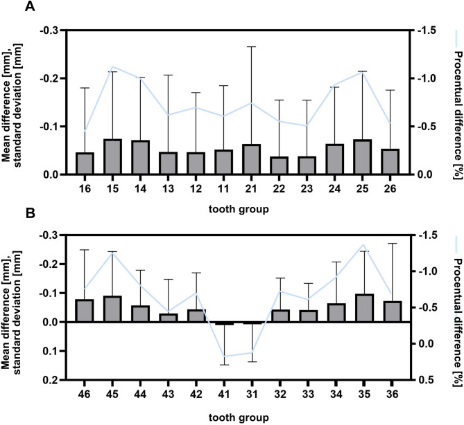
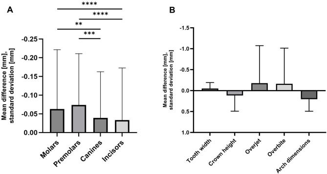
Results: Differences of up to 0.3 mm were observed between manual and partially automated digital measurements for sagittal, transversal and vertical parameters. Teeth with close proximal contact to adjacent teeth and teeth in jaws with a negative space analysis result showed an increased difference between manual and partially automated digital measurements, although this was not clinically relevant. The time required to perform digital measurements was statistically significantly reduced.
Conclusions: Partially automated digital impression analysis offers an accurate, highly efficient and time-saving alternative to traditional manual impression analysis.
期刊介绍:
Head & Face Medicine is a multidisciplinary open access journal that publishes basic and clinical research concerning all aspects of cranial, facial and oral conditions.
The journal covers all aspects of cranial, facial and oral diseases and their management. It has been designed as a multidisciplinary journal for clinicians and researchers involved in the diagnostic and therapeutic aspects of diseases which affect the human head and face. The journal is wide-ranging, covering the development, aetiology, epidemiology and therapy of head and face diseases to the basic science that underlies these diseases. Management of head and face diseases includes all aspects of surgical and non-surgical treatments including psychopharmacological therapies.
 求助内容:
求助内容: 应助结果提醒方式:


