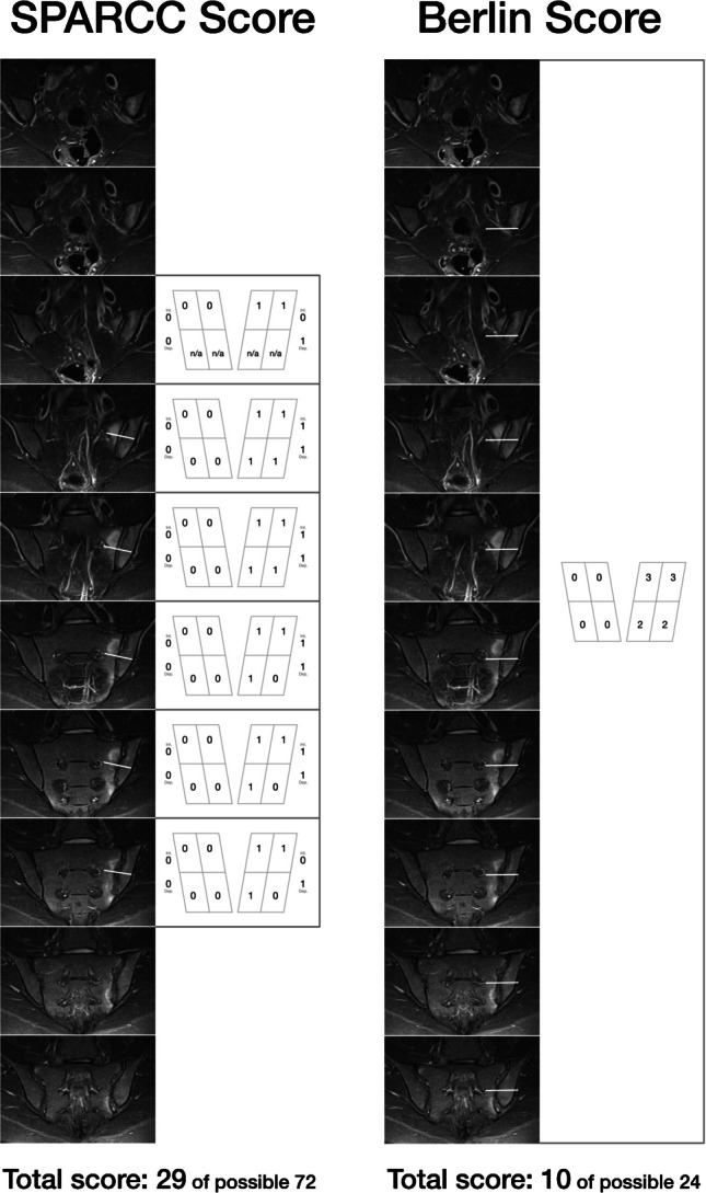
{"title":"Imaging in clinical trials of axial spondyloarthritis: what type of imaging should be used.","authors":"Iris Eshed, Kay Geert A Hermann","doi":"10.1007/s00256-025-04935-0","DOIUrl":null,"url":null,"abstract":"<p><p>Axial spondyloarthritis (axSpA) is a chronic inflammatory condition predominantly affecting the sacroiliac joints and spine. Early and accurate diagnosis is crucial to prevent structural damage and improve patient outcomes. Imaging plays a pivotal role in axSpA diagnosis, monitoring, and clinical trials, offering insights into both inflammatory activity and structural progression. Conventional radiography has been foundational for detecting structural changes, such as syndesmophytes and erosions, but it is limited by poor sensitivity for early disease detection and significant interobserver variability. Advanced imaging modalities, such as magnetic resonance imaging (MRI) and low-dose computed tomography (ld-CT), have emerged as more sensitive tools. MRI excels in identifying active inflammation, particularly bone marrow edema, and is integral to early diagnosis and disease monitoring. ld-CT provides superior spatial resolution for detecting structural lesions while minimizing radiation exposure. However, challenges remain in achieving standardized imaging protocols and consistent scoring systems across clinical trials. Scoring systems like the modified Stoke Ankylosing Spondylitis Spine Score (mSASSS), Spondyloarthritis Research Consortium of Canada (SPARCC) scores, and Berlin methods require rigorous calibration to ensure reliability. The purpose of this review is to explore the strengths and limitations as well as the use in clinical trials of the different imaging modalities and to offer guidance on selecting the most suitable imaging techniques for assessing both disease activity and structural progression in clinical trials.</p>","PeriodicalId":21783,"journal":{"name":"Skeletal Radiology","volume":" ","pages":"2399-2409"},"PeriodicalIF":2.2000,"publicationDate":"2025-11-01","publicationTypes":"Journal Article","fieldsOfStudy":null,"isOpenAccess":false,"openAccessPdf":"https://www.ncbi.nlm.nih.gov/pmc/articles/PMC12460507/pdf/","citationCount":"0","resultStr":null,"platform":"Semanticscholar","paperid":null,"PeriodicalName":"Skeletal Radiology","FirstCategoryId":"3","ListUrlMain":"https://doi.org/10.1007/s00256-025-04935-0","RegionNum":3,"RegionCategory":"医学","ArticlePicture":[],"TitleCN":null,"AbstractTextCN":null,"PMCID":null,"EPubDate":"2025/5/10 0:00:00","PubModel":"Epub","JCR":"Q2","JCRName":"ORTHOPEDICS","Score":null,"Total":0}
引用次数: 0
Abstract
Axial spondyloarthritis (axSpA) is a chronic inflammatory condition predominantly affecting the sacroiliac joints and spine. Early and accurate diagnosis is crucial to prevent structural damage and improve patient outcomes. Imaging plays a pivotal role in axSpA diagnosis, monitoring, and clinical trials, offering insights into both inflammatory activity and structural progression. Conventional radiography has been foundational for detecting structural changes, such as syndesmophytes and erosions, but it is limited by poor sensitivity for early disease detection and significant interobserver variability. Advanced imaging modalities, such as magnetic resonance imaging (MRI) and low-dose computed tomography (ld-CT), have emerged as more sensitive tools. MRI excels in identifying active inflammation, particularly bone marrow edema, and is integral to early diagnosis and disease monitoring. ld-CT provides superior spatial resolution for detecting structural lesions while minimizing radiation exposure. However, challenges remain in achieving standardized imaging protocols and consistent scoring systems across clinical trials. Scoring systems like the modified Stoke Ankylosing Spondylitis Spine Score (mSASSS), Spondyloarthritis Research Consortium of Canada (SPARCC) scores, and Berlin methods require rigorous calibration to ensure reliability. The purpose of this review is to explore the strengths and limitations as well as the use in clinical trials of the different imaging modalities and to offer guidance on selecting the most suitable imaging techniques for assessing both disease activity and structural progression in clinical trials.
期刊介绍:
Skeletal Radiology provides a forum for the dissemination of current knowledge and information dealing with disorders of the musculoskeletal system including the spine. While emphasizing the radiological aspects of the many varied skeletal abnormalities, the journal also adopts an interdisciplinary approach, reflecting the membership of the International Skeletal Society. Thus, the anatomical, pathological, physiological, clinical, metabolic and epidemiological aspects of the many entities affecting the skeleton receive appropriate consideration.
This is the Journal of the International Skeletal Society and the Official Journal of the Society of Skeletal Radiology and the Australasian Musculoskelelal Imaging Group.

 求助内容:
求助内容: 应助结果提醒方式:


